
 Türkçe
 Deutsch
 العربية
 Русский

Heart valve diseases that develop later in children are cardiac pathologies that occur not at birth but in later stages of life and progress with structural or functional deterioration of the heart valves. The most common cause is acute rheumatic fever (ARF), in which the immune response after infection leads to damage in the valve tissue.
Rheumatic heart valve disease in children is especially seen following streptococcal infections. The mitral valve is the most frequently affected structure, and in the early stage it causes valve insufficiency, while in later stages it may present clinically as both stenosis and insufficiency. Early diagnosis and appropriate antibiotic treatment reduce disease progression.
Symptoms of acquired heart valve insufficiency may include easy fatigue, shortness of breath, palpitations, and growth retardation. Detection of a murmur during physical examination is important for diagnosis. Echocardiography is the primary imaging method used to evaluate valve functions and determine the severity of the disease.
Treatment of heart valve disease in children is planned according to the type and severity of valve damage. In mild cases, regular follow-up may be sufficient, while in advanced cases medical treatment or surgical intervention may be required. Long-term monitoring is critically important for preventing complications.
| Things You Should Know | Information |
| Definition | Heart valve diseases that develop later in children are conditions that occur due to structural or functional deterioration of the heart valves, not congenital, and associated with infections, rheumatic diseases, connective tissue diseases, or other systemic conditions. |
| Most Common Cause | In developing countries, the most common cause is rheumatic heart disease due to acute rheumatic fever. Apart from this, infective endocarditis and Kawasaki disease are among other causes. In addition, systemic rheumatologic diseases, other autoimmune diseases or metabolic diseases may also lead to valve damage. |
| Affected Valves | The mitral valve is most frequently affected. The aortic valve is the second most commonly involved. More rarely, the tricuspid and pulmonary valves may be affected. |
| Pathophysiology | Thickening, fibrosis, calcification, or vegetation may develop in the valve leaflets. This condition leads to stenosis or insufficiency (regurgitation) of the valve, causing pressure changes within the heart and enlargement of the heart chambers. |
| Risk Factors | Untreated streptococcal infections, inadequate antibiotic use, immune system diseases, history of heart surgery, long-term presence of central catheters, and intravenous drug use are risk factors. |
| Symptoms | Shortness of breath, easy fatigue, palpitations, chest pain, fainting, growth retardation, exercise intolerance, and swelling in the feet in advanced cases may occur. Some children may remain asymptomatic for a long time. |
| Physical Examination Findings | The most important finding is the presence of a murmur. Additionally, cardiomegaly, tachycardia, peripheral edema, hepatomegaly, and cyanosis may be detected. |
| Diagnostic Methods | Echocardiography is the main diagnostic method and shows valve structure and function in detail. Electrocardiography (ECG), chest radiography, and when necessary cardiac MRI/CT, PET, and scintigraphy are other supportive examinations. |
| Possible Complications | Heart failure, pulmonary hypertension, arrhythmia, thromboembolism, and infective endocarditis may develop. In the long term, severe valve damage may require surgery. |
| Treatment Approaches | Treatment is planned according to the underlying cause. Antibiotic therapy (for rheumatic fever or infective endocarditis), anti-inflammatory medications, diuretics, and other heart failure treatments may be applied. |
| Interventional and Surgical Treatment | In cases of severe valve stenosis or insufficiency, balloon valvuloplasty, valve repair, or valve replacement may be required. The surgical decision is made according to the degree of valve damage and clinical findings. |
| Follow-up | Regular cardiology control and periodic echocardiography are necessary. In rheumatic heart disease, long-term antibiotic prophylaxis may be applied. |
| Prevention | Early and appropriate antibiotic treatment of streptococcal throat infections is the most effective method for preventing rheumatic heart disease. Infective endocarditis prophylaxis may be recommended in high-risk patients. |
| Prognosis | It depends on the severity of the disease and early diagnosis and treatment. While quality of life can be preserved in mild cases, advanced cases may require surgical intervention and long-term follow-up. |

PROF. DR.
İbrahim Cansaran TANIDIR
Pediatric Cardiology Specialist
İbrahim Cansaran TANIDIR
Pediatric Cardiology Specialist
Prof. Dr. İbrahim Cansaran Tanıdır is an internationally recognized expert with nearly 20 years of clinical, academic, and interventional experience in congenital heart diseases, pediatric interventional cardiology, hybrid procedures, and radiation-reduced / fluoroscopy-free catheterization techniques.
To date, he has performed more than 10,000 pediatric cardiac catheterization procedures both in our country and abroad; and has pioneered advanced interventional and hybrid applications in complex structural heart diseases in our country. His academic productivity represents a strong scientific authority with 110 SCI/SCIE publications, 1,440 citations, and an H-index of 18 (Google Scholar, 2025).
If my child’s heart was healthy at birth, how can it become diseased later?
As a parent, it is completely natural to have difficulty understanding how your child’s heart, which was perfectly healthy at birth, can become diseased later. The human body is not a closed box; it constantly interacts with its environment, microbes, and various triggers. At the core of heart diseases that develop later is usually a story of a “misunderstanding.” Our body’s defense system, the immune system, is responsible for protecting the child against microbes coming from outside. However, sometimes while trying to destroy the microbe, this system may mistake its own tissues, which closely resemble the structure of the microbe, as enemies.
The most common example of this situation is Acute Rheumatic Fever, known among the public as “Heart Rheumatism.” Here, the culprit is not a microbe directly entering the heart but the uncontrolled immune response triggered by the microbe. Another scenario is when bacteria leak into the bloodstream and directly settle on the heart valves, which we call “Infective Endocarditis.” In both cases, the result is the deterioration, thickening, or calcification of the heart’s delicate and perfectly functioning valve structure. This process sometimes progresses very quietly and insidiously, and sometimes appears suddenly with a very dramatic clinical picture. The important thing is to know that this process is preventable or can be stopped at an early stage.
How does the “beta microbe” and throat infection affect the heart?
The “beta microbe” you frequently hear about during winter months or school periods is actually a bacterium called Group A Beta Hemolytic Streptococcus. This bacterium settles in the tonsils and causes a throat infection accompanied by fever and difficulty swallowing. Most of the time, this infection heals without problems with appropriate antibiotic treatment. However, if it is not treated or treated inadequately, complications may arise.
Some proteins on the outer surface of the beta microbe are surprisingly very similar to proteins in the valve tissue and joints of the human heart. Your child’s immune system produces soldiers called antibodies to kill the microbe. These soldiers eliminate the microbe but cannot stop the war. Thinking “it resembles the enemy,” they continue to attack the child’s own perfectly healthy mitral or aortic valve. In medicine, we call this “molecular mimicry.”
As a result of this attack, inflammation begins in the valves. The valve leaflets swell and small clots form on them. Over time, as this inflammation heals, it leaves behind a hard, non-elastic tissue we call “scar tissue.” As a result, the valve leaflets either stick to each other and have difficulty opening (stenosis) or shrink and cannot close completely (insufficiency). The heart becomes tired and enlarged while trying to pump blood through a narrow gate or while dealing with a valve that constantly leaks backward. This chain reaction explains how a simple sore throat can affect the heart.
What symptoms are seen in a child who has had Acute Rheumatic Fever?
Rheumatic fever usually occurs 2 to 4 weeks after a throat infection. In other words, your child has recovered from the sore throat and you think everything is fine, but after a while completely different complaints begin. This disease can involve many parts of the body and the symptoms may vary in severity in each child.
The most common and prominent symptom is seen in the joints. The characteristics of joint involvement are as follows:
- Severe pain
- Swelling
- Redness
- Increase in temperature
- Restriction of movement
This joint pain has a very typical characteristic: it is “migratory.” For example, your child’s right knee becomes swollen and painful. A day or two later it heals completely without leaving any trace, but this time the left ankle becomes swollen. The pain may sometimes be so severe that the child cannot step on it, and may even be unable to tolerate a blanket touching it.
Another important symptom is involvement of the nervous system. In this condition called “Sydenham’s Chorea,” the movement centers in the brain are affected. Your child may begin to show clumsiness that was not present before. They may drop the glass in their hand, their handwriting may deteriorate, and they may be unable to button their clothes. In more advanced cases, involuntary sudden movements of the arms and legs and facial grimacing may occur. This condition may sometimes be mistaken for “spoiled behavior” or a “tic.” Emotional fluctuations, unexplained crying, or laughing may also accompany it.
However, the most critical involvement for us is heart involvement (Carditis). Because the joints hurt, the family brings the child to the doctor quickly, but heart inflammation does not cause pain. It progresses silently. If your child experiences easy fatigue, shortness of breath, or palpitations, or if a “murmur” that was not previously present is heard in the heart during a doctor’s examination, we suspect heart involvement.
What tests and criteria do doctors look at when making a diagnosis?
Diagnosing rheumatic fever is like assembling the pieces of a puzzle. It is not possible to say “yes, this child is sick” with a single blood test. As doctors, we use an international guideline called the “Jones Criteria.”
First, we must prove that the child has recently had a beta infection. The tests used for this are:
- Throat culture
- Rapid antigen test
- ASO (Antistreptolysin O) value
A high ASO value shows that the child has recently encountered this microbe. However, an elevated ASO alone does not establish the diagnosis of the disease; it is only a clue.
In addition, we examine blood values that show general inflammation in the body.
- CRP and
- High sedimentation (ESR) values indicate that there is an active battle in the body.
The most critical stage of diagnosis is imaging of the heart. The device called echocardiography (ECHO) allows us to see inside the heart using sound waves. With ECHO, we examine the following in detail:
- Valve structure
- Valve thickening
- Valve leaks
- Strength of the heart muscle
- Fluid around the heart
Sometimes even if no murmur is heard from the outside, we can detect leakage in the valves at a very early stage thanks to ECHO. This allows us to start treatment early.
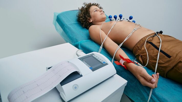
- ECG contains typical findings for patients in whom echocardiography findings are not detected or are borderline.
Contact us now to get detailed information about treatments and to create an appointment!
Why is the use of aspirin and cortisone necessary during the treatment process?
After making the diagnosis, we start treatment without delay. The treatment has three main goals:
- To completely eliminate the microbe in the body,
- To extinguish the inflammation caused by the immune system and
- To protect the heart.
The first step is antibiotics. We usually administer a penicillin group antibiotic so that no beta microbe remains in the child’s throat or body. This is intended to eliminate the source that triggered the disease.
The second step is suppressing inflammation. Here, the choice of medication changes depending on which organ is affected by the disease. If there is only swelling and pain in the joints, we start high-dose Aspirin (salicylate) treatment. Normally, we do not prefer to use aspirin in children because it may be confused with some viral infections and strain the liver. However, rheumatic fever is one of the rare diseases in which aspirin is life-saving and safely used in children. Aspirin relieves joint pain and swelling with a miraculous speed, usually completely within 24–48 hours. This rapid response is itself a sign confirming the diagnosis for us.
If there is inflammation in the heart (carditis), the situation is more serious and aspirin alone may be insufficient. To minimize damage to the heart valves, we need a stronger anti-inflammatory agent: cortisone (prednisone). Cortisone suppresses the excessive immune attack on the heart. It is usually used for about 2–4 weeks and then gradually reduced and discontinued. During this period, we recommend a salt-free diet to counteract the salt-retaining effect of cortisone in the body. Our aim is to prevent that severe inflammation in the acute period from turning into permanent scar tissue (sequela).
What is infective endocarditis and how does it destroy the heart valve?
Infective endocarditis develops through a completely different mechanism than rheumatic fever. In rheumatic fever the immune system attacks the heart, whereas in endocarditis the bacteria themselves settle on the heart valves. You can think of this as a nest of microbes being established inside the heart.
This disease is usually seen in children who already have a predisposition in the heart. Those with congenital heart defects, children who previously had rheumatic valve disease, or those with artificial heart valves are at risk. However, it can rarely occur even in a completely healthy heart.
Bacteria in the bloodstream adhere to the damaged or rough surface of the heart valve. There they form a protected structure surrounded by blood cells and clots. In medical terms we call this “vegetation.” This structure protects bacteria from antibiotics and the body’s defense cells. The bacteria multiply rapidly here and begin to literally eat away at the valve. They can perforate the valve, destroy it, or form abscesses around the valve.
The most dangerous aspect of infective endocarditis is not only that it destroys the heart. Pieces that break off from these clusters of microbes called vegetation can spread throughout the body through the bloodstream. They can travel to the brain and cause stroke, to the kidneys and cause failure, or to the lungs and cause vascular blockages. Therefore, once diagnosed, hospitalization and long-term high-dose intravenous antibiotic treatment (4–8 weeks) and in some patients surgical intervention are required.
What is the relationship between dental health, oral hygiene, and the heart?
This connection is one of the most surprising yet vital issues for parents. The oral cavity is one of the areas in our body that contains the highest concentration of bacteria. In a healthy mouth and dental structure, these bacteria remain in place and do not cause problems. However, during tooth decay, gum diseases (gingivitis), or bleeding procedures such as tooth extraction, these bacteria in the mouth can easily mix into the bloodstream. In medicine we call this “bacteremia.”
In a healthy person, the immune system clears these bacteria in the bloodstream within minutes. However, in children who already have damage to a heart valve, the situation is different. The bacteria that enter the bloodstream love to attach to that damaged heart valve. There they settle and multiply and initiate the deadly condition called Infective Endocarditis that we mentioned earlier.
For this reason, brushing teeth is just as important as taking heart medications for a child with heart disease. Decayed teeth are like a time bomb for the heart and must be treated.
In addition, there is a preventive measure called “Endocarditis Prophylaxis.” Patients in the high-risk group (those with prosthetic valves, those who have previously had endocarditis, and some congenital heart patients) must take antibiotics before undergoing dental treatment or any oral procedure that may cause bleeding. A single dose of antibiotic taken 30–60 minutes before the procedure eliminates bacteria that may enter the bloodstream before they reach the heart. This simple precaution is life-saving.
What are the balloon or surgical options in the treatment of valve disease?
When severe stenosis or insufficiency develops in a heart valve, medications may no longer be sufficient and a mechanical correction may be required. Fortunately, today it is not necessary to open the chest for every case.
If the problem is valve stenosis, especially in the mitral and pulmonary valves, the method called “Balloon Valvuloplasty” is very successful. This procedure is performed in an angiography laboratory rather than an operating room.
A thin sheath (catheter) is inserted through the vessel in the child’s groin.
- A deflated balloon is advanced into the narrowed valve.
- The balloon is inflated with pressure applied from outside.
- The force of the balloon tears apart the valve leaflets that have stuck together and relieves the stenosis.
- With this method, the child can be discharged the next day without any incision scar on the chest and breathe comfortably for many years.
However, if the structure of the valve is not suitable for balloon treatment or if there is severe leakage (insufficiency), surgery becomes necessary. In pediatric cardiac surgery, our basic philosophy is “Repair the Valve, Do Not Replace It.” This is because the child’s own valve is living tissue and grows together with the child. It also does not require blood-thinning medication.
If repair is not possible, valve replacement (replacement) is performed. There are two options: metal (mechanical) valves and biological valves. Metal valves are very durable and function for life. However, because they carry a risk of blood clotting, they require the child to use a strong blood thinner such as “Warfarin” for life. Children using this medication must pay close attention to their diet (foods containing vitamin K such as green vegetables) and have blood tests every month. Biological valves are made from animal tissues and do not require blood thinners, but they may calcify and deteriorate quickly in children, which means another surgery. Which method will be chosen is decided after detailed discussion according to the child’s age, the family’s social circumstances, and the condition of the valve.
Contact us now to get detailed information about treatments and to create an appointment!
Why does my child have to receive protective penicillin injections for years?
For a child who has had rheumatic heart disease and their family, perhaps the most challenging process is the injection treatment that will last for years. However, these injections are the most critical insurance of the treatment. The worst feature of rheumatic fever is that it is recurrent. Each time your child contracts the beta microbe again, the immune system flares up again and increases the damage to the heart valve even more. A valve that survived the first attack with mild leakage may completely fail in the second attack.
To reduce this risk to zero, we apply “Secondary Prophylaxis.” The most effective method for this is the “Benzathine Penicillin G” injection administered in the hip every 21 days (once every 3 weeks). This injection slowly mixes into the bloodstream from the muscle tissue and creates a continuous shield in the body. The moment the beta microbe tries to settle in the throat, the ready antibiotic in the blood eliminates it.
The duration of injections varies depending on the patient’s condition:
- If there was only joint involvement and no heart damage: Usually until age 21 or for 5 years after the last attack.
- If there was heart involvement but it healed: Usually for 10 years or until age 21.
- If permanent valve damage remains in the heart or if valve surgery has been performed: At least until age 40, sometimes for life.
Penicillin tablets taken orally unfortunately do not provide protection as reliable as injections. They may be forgotten or poorly absorbed. Therefore, giving up the injection by saying “it hurts a little” means putting the child’s heart at great risk.
Which “Red Flag” symptoms require urgent medical attention?
While your child’s routine follow-ups continue, some situations you observe at home may indicate that the heart is under strain or that something is going wrong. When you notice these symptoms, you should consult a doctor without waiting for the next scheduled check-up.
The main situations you should pay attention to are:
- Excessive sweating during feeding
- Stopping or slowing of weight gain
- Easy fatigue with exertion
- Bluish discoloration of the lips or nail beds
- Waking up at night with shortness of breath
- Desire to sleep with a high pillow
- Swelling in the eyelids or legs
- Unexplained fainting episodes
- Complaint of chest pain
- Persistent fever that does not subside
- Heartbeats being so fast that they can be noticed from outside
- Presence of other accompanying findings (persistent fever, joint pain, swelling in joints…)
Can children with heart valve disease live a normal life?
The answer to this question is a big “Yes.” Being diagnosed with heart valve disease does not mean that your child’s life is over or that they must constantly stay in bed. On the contrary, with proper treatment and regular follow-up, the vast majority of these children live completely normal, active, and productive lives. They can go to school, have a profession, get married, and have children.
The important thing here is to know the limits imposed by the disease and live accordingly. For example, it may be inconvenient for a child with severe valve stenosis or who uses blood thinners to engage in professional-level, highly competitive sports such as football, basketball, or boxing. Those using blood thinners should avoid trauma. However, activities such as light-paced walking and swimming are usually allowed and are even recommended for heart health. Because the heart is also a muscle and needs conditioning:
The child’s psychology is also very important in this process. Treating them as a “sick child,” being overly protective, may damage the child’s self-confidence. It is necessary to explain the illness in language appropriate to their age, explain why medications are necessary, and not isolate them from social life.
Frequently Asked Questions
After which infections can Heart Valve Diseases Developing Later in Children occur?
In childhood, acute rheumatic fever developing especially due to beta hemolytic streptococcal infection may cause damage to the heart valves. Rarely, severe infections such as bacterial endocarditis may also lead to permanent deterioration in the valve tissue.
How do Heart Valve Diseases Developing Later in Children affect heart functions?
When narrowing or insufficiency occurs in the valves, blood flow is disrupted and the heart has to work harder. Over time, this condition may lead to thickening of the heart muscle, enlargement, and the risk of heart failure in advanced stages.
With which symptoms do Heart Valve Diseases Developing Later in Children manifest?
Easy fatigue, shortness of breath, palpitations, chest pain, and fainting episodes are common symptoms. In young children, failure to gain weight and feeding difficulties may also be indirect signs of valve disease.
In which age group are Heart Valve Diseases Developing Later in Children more common?
They are most frequently seen in school-age children and adolescents. The reason for this is that rheumatic fever is more common in this age group. However, valve damage does not develop in every child who has had an infection. Apart from this, while Kawasaki syndrome is common in childhood, heart valve diseases related to systemic diseases may develop over time.
If Heart Valve Diseases Developing Later in Children progress, which complications may occur?
If left untreated, heart failure, rhythm disorders, increased pulmonary pressure, and rarely clot formation may occur. In advanced cases, quality of life may decrease significantly and surgical intervention may be required.
How are Heart Valve Diseases Developing Later in Children diagnosed?
Hearing a murmur during physical examination raises suspicion. Diagnosis is usually made with echocardiography. This method allows detailed evaluation of the structure of the valves, blood flow, and the condition of the heart chambers. After a definite diagnosis/pre-diagnosis is made, further examinations may be performed.
Which methods are applied in the treatment of Heart Valve Diseases Developing Later in Children?
Treatment is planned according to the degree of valve damage. Drug therapy aims to reduce the workload of the heart. In advanced cases, surgical methods such as valve repair or valve replacement may come to the agenda.
What is the recovery process after surgery for Heart Valve Diseases Developing Later in Children?
After surgery, children can usually return home within a few weeks. Returning to daily activities will vary depending on the surgical procedure performed. However, regular cardiology check-ups are important. Long-term medication use may be required in patients who undergo valve replacement.
Do Heart Valve Diseases Developing Later in Children prevent sports activities?
Sports restrictions may be necessary depending on the severity of the disease. While controlled exercise may be recommended in mild cases, heavy and competitive sports should be avoided in advanced valve disease.
How can Heart Valve Diseases Developing Later in Children be prevented?
Early diagnosis of throat infections and treatment with appropriate antibiotics reduces the risk of rheumatic fever. In diagnosed children, regular follow-up and preventive antibiotic applications can prevent the progression of valve damage. In heart involvement of systemic diseases, close monitoring and medications initiated depending on disease progression may delay heart involvement.
Blog Posts

Exercise Stress Test in Children: Purpose, Procedure and Results
Exercise stress testing in children is [...]
Read More
29
Sep
Sep
Holter ECG Monitoring in Children: Procedure, Uses and Results
Holter ECG in children is a [...]
Read More
29
Sep
Sep

Pediatric Echocardiography (ECHO): Procedure, Uses and Results
Pediatric echocardiography is a painless ultrasound [...]
Read More
29
Sep
Sep
Pediatric ECG (Electrocardiogram): Procedure, Uses and Results
Pediatric ECG (Electrocardiography) is a completely [...]
Read More
29
Sep
Sep

Pulmonary Valve Stenosis: Symptoms, Causes, Diagnosis and Treatment
Pulmonary valve stenosis (pulmonary stenosis) is [...]
Read More
29
Sep
Sep
Ventricular Septal Defect (VSD): Symptoms, Causes and Treatment Options
Ventricular septal defect (VSD) is a [...]
Read More
29
Sep
Sep
Atrial Septal Defect (ASD): Symptoms, Causes and Treatment Options
Atrial septal defect (ASD) is a [...]
Read More
29
Sep
Sep

Shortness of Breath in Children: Causes, Symptoms and Treatment Options
Shortness of breath in children is [...]
Read More
10
Aug
Aug