
 Türkçe
 Deutsch
 العربية
 Русский

Congenital heart diseases in children are cardiac anomalies that arise due to structural developmental defects of the heart and are present from birth. Among the most common forms are holes in the heart (atrial septal defect (ASD), ventricular septal defect (VSD)), patent ductus arteriosus (PDA), valve stenosis, and valve insufficiency; these conditions can affect blood flow and oxygenation.
Symptoms of a hole in the heart (ASD and VSD) may appear as rapid breathing, bluish discoloration, failure to gain weight, and frequent infections. Septal defects cause abnormal blood passage between the right and left chambers of the heart. While small defects require follow-up, large defects may require surgery or catheter-based intervention.
Findings of valve stenosis and valve regurgitation are associated with a murmur, easy fatigue, chest pain, and fainting. In aortic or pulmonary valve stenosis, forward blood flow is restricted; in valve insufficiency, backward leakage occurs. Diagnosis is made by echocardiography, and treatment is planned according to the severity.
The diagnostic and treatment process in congenital heart diseases includes fetal echocardiography, newborn screenings, and pediatric cardiology evaluation. Early diagnosis is critical for preventing complications. Follow-up encompasses monitoring, medical treatment, interventional methods, or surgical intervention options.
| What You Need to Know | Information |
| Definition | Congenital heart diseases in children are structural abnormalities of the heart or great vessels that occur during development in the womb. Holes between the heart chambers (ASD, VSD), valve stenosis or insufficiency, and great vessel anomalies are the most common forms. |
| Incidence | Congenital heart disease is seen in approximately 0.8–1% of live births. Mild forms may sometimes be diagnosed in childhood, while some cases may be diagnosed in adulthood. |
| Causes | In most cases, the exact cause is unknown. Genetic factors, chromosomal anomalies (for example Down syndrome), family history, maternal infections during pregnancy, diabetes, certain medications, or exposure to harmful substances are among the risk factors. |
| Classification | In general, they are divided into two groups: cyanotic (accompanied by bluish discoloration) and acyanotic (without bluish discoloration) heart diseases. They can also be classified as simple (isolated small defects) and complex (forms involving multiple structural abnormalities). |
| Most Common Defects | Atrial septal defect (ASD), ventricular septal defect (VSD), patent ductus arteriosus (PDA), pulmonary valve stenosis, aortic stenosis, and tetralogy of Fallot are the most common congenital heart diseases. |
| Hole (Septal Defect) | This is the presence of an opening in the wall between the atria (ASD) or ventricles (VSD) of the heart. While small defects may not cause symptoms, large defects can lead to heart failure and growth retardation. |
| Patent ductus arteriosus (PDA) | PDA is an opening between the aorta and the pulmonary artery that exists in all babies in the womb and enables survival. After birth, the PDA closes rapidly, first functionally and then anatomically. If it does not close, it may lead to findings/complaints depending on its size. |
| Valve Stenosis | This is the restriction of blood flow due to the heart valves not opening fully. Pulmonary and aortic valve stenosis are the most common. Severe stenosis causes an increased workload on the heart muscle. |
| Valve Insufficiency (Regurgitation) | This is the backward leakage of blood due to the heart valves not closing completely. Over time, enlargement of the heart chambers and functional impairment may develop. |
| Symptoms | Rapid breathing, easy fatigue, difficulty feeding, failure to gain weight, frequent respiratory tract infections, bluish discoloration (especially on the lips and nails), sweating, and fainting may be seen. Mild cases may remain asymptomatic for a long time. |
| Physical Examination Findings | A heart murmur, pulse differences, bluish discoloration, respiratory distress, and growth retardation can be detected on physical examination. |
| Diagnostic Methods | Echocardiography (ECHO) is the primary diagnostic method. Electrocardiography (ECG), chest X-ray, cardiac MRI, and in some cases cardiac catheterization support the diagnosis. |
| Treatment Options | Follow-up, medical therapy (heart failure treatment), catheter-based interventional procedures (hole closure with a device, balloon valvuloplasty), or open-heart surgery may be performed. Treatment is planned according to the type of defect, the patient’s age, weight, and severity. |
| Surgical Intervention | Surgical correction may be required in large septal defects, severe valve anomalies, or complex heart diseases. Regular follow-up after surgery is important. |
| Complications | If left untreated, heart failure, pulmonary hypertension, arrhythmias, infective endocarditis, and growth retardation may develop. |
| Monitoring and Follow-up | Patients should be monitored at regular intervals by pediatric cardiology. Some small defects may close spontaneously or mild stenosis may resolve on its own, but some require lifelong follow-up. |
| Prognosis | With early diagnosis and appropriate treatment, many children can live a normal life. Prognosis depends on the type and severity of the disease. |
| Prevention | Although not all cases can be prevented, pre-pregnancy genetic counseling, regular follow-up during pregnancy, avoiding harmful substances, and controlling chronic diseases can reduce risk. |

PROF. DR.
İbrahim Cansaran TANIDIR
Pediatric Cardiology Specialist
İbrahim Cansaran TANIDIR
Pediatric Cardiology Specialist
Prof. Dr. İbrahim Cansaran Tanıdır is an internationally recognized expert with nearly 20 years of clinical, academic, and interventional experience in congenital heart diseases, pediatric interventional cardiology, hybrid procedures, and radiation-reduced / fluoroscopy-free catheterization techniques.
To date, he has performed more than 10,000 pediatric cardiac catheterization procedures both in our country and abroad; and has pioneered advanced interventional and hybrid applications in complex structural heart diseases in our country. His academic productivity represents a strong scientific authority with 110 SCI/SCIE publications, 1,440 citations, and an H-index of 18 (Google Scholar, 2025).
Why Do Congenital Heart Diseases Occur, and Should Families Blame Themselves?
In the eyes of every mother and father who walks through the clinic door and is told their child has a heart disease, we see the same concern and the same question: “Where did we go wrong?” or “Was it something I ate while I was pregnant that caused this?”
First of all, you should keep your mind at ease; the vast majority of congenital heart diseases are not due to a single cause and do not result from simple things you did or did not do. In medicine, we call this a “multifactorial” process; that is, there is a complex interaction between genetic predisposition and environmental factors.
The formation of the heart is completed very early in pregnancy, between the 3rd and 8th weeks, when most expectant mothers have only just learned they are pregnant. In this sensitive period, small errors in genetic codes or external factors can alter the architecture of the heart. Although having a congenital heart disease in the family—meaning in the mother, father, or siblings—slightly increases the risk, the vast majority of babies born with heart disease do not have a known family history of heart disease. Therefore, genetic inheritance is not always the single determining factor.
However, some conditions have been scientifically proven to increase the risk. For example, if the mother has diabetes (high blood sugar) before or during pregnancy and blood sugar is poorly controlled, it can affect the baby’s heart development. Similarly, severe viral infections such as rubella in the first trimester of pregnancy, or the use of certain potent medications without medical supervision, are also among the risk factors. Consanguineous marriages can also increase the frequency of some genetically inherited syndromes and, consequently, heart diseases. Despite all this, we can also encounter this condition in the babies of mothers who carry no risk factors and have had an extremely healthy pregnancy. Therefore, instead of questioning the past and blaming yourself, focusing on solving the current situation is the best thing you can do for your child.
What Symptoms Suggest Heart Disease in Babies?
A newborn baby cannot explain their problem or show their pain. Therefore, their language is the reactions their bodies give. While heart diseases can sometimes present with a very dramatic picture—such as bluish discoloration or shock immediately after birth—they can also progress very silently. Especially large defects or vessel patency that lead to heart failure signal their presence in babies with typical feeding problems.
For a baby, sucking is a task that requires serious effort, like an adult running on a treadmill. A baby with a heart problem who has excess blood flow to the lungs cannot tolerate this effort. While breastfeeding or bottle-feeding, they tire very quickly, frequently release the nipple to breathe, and want to rest. During this, the baby’s metabolism speeds up due to the exertion. Another finding that families most frequently report to us and that we take very seriously is “sweating.” Especially if beads of sweat accumulate on the baby’s forehead during feeding, this may indicate that the heart is working beyond its capacity to pump blood to the body.
Some basic symptoms you should watch for in infancy are:
- Tiring quickly while feeding
- Stagnation in weight gain
- Rapid breathing
- Excessive sweating in the forehead area
- Frequent recurrent lung infections
- Swelling of the eyelids
- Restlessness
- Making groaning-like sounds
- A fast heartbeat
These symptoms alone do not establish a diagnosis of heart disease, but they do indicate that specialist evaluation is definitely needed. In many patients who come in saying, “My baby is not gaining weight and has no appetite,” we can see that the underlying reason is not metabolic, but increased energy consumption due to the heart working harder.
Which Complaints in Older Children May Be Signs of Heart Disease?
In some children who pass infancy without problems or whose symptoms are overlooked, heart disease may become apparent at school age or during adolescence. Since children in this age group can express themselves, the process works a bit differently. Families or teachers usually notice a decline in the child’s performance during physical activities.
For example, in a child who gets tired before everyone else while playing ball with friends, feels the need to leave the game and squat to rest, or becomes much more short of breath than peers while climbing stairs, heart reserves should be evaluated. This limitation in exercise capacity may be a sign that the heart is struggling to meet the body’s increasing oxygen demand.
Possible complaints that may be seen in school-age children include:
- Chest pain with exercise
- A feeling of palpitations
- Fainting
- Easy fatigue, tiring earlier than peers
- Dizziness, dimming of vision
- Swelling in the legs
- Shortness of breath
Here, it is necessary to open a special parenthesis regarding “chest pain.” The number of families presenting to pediatric clinics with a complaint of chest pain is quite high. However, the good news is that the vast majority of chest pains in children are not caused by the heart. Muscle pain, growth pains, reflux, or stress-related pains are usually more prominent. Nevertheless, chest pain that occurs especially during exertion (while running, swimming) and stops the child, or fainting episodes, are conditions that should not be neglected and require serious cardiac evaluation.
Contact us now to get detailed information about treatments and to create an appointment!
What Does Hearing a “Murmur” on Examination Mean?
During a routine check-up, after your doctor places the stethoscope on your child’s chest and says, “I hear a sound, a murmur in the heart,” it can be a very frightening moment for parents. But stay calm, because a murmur does not always mean disease. A murmur is only a sound; it is the hum made by blood flowing inside the heart.
We can explain this with the example of a garden hose. If you slightly squeeze the end of a hose with water flowing through it, or greatly increase the flow speed, the sound changes and you hear a “hissing” sound. The same is true in the heart. If blood is trying to pass through a narrowed valve, or if it is spurting under pressure from one side to the other through a hole in the heart, it creates a pathological sound. However, sometimes a sound can be heard even when the heart’s structure is completely normal, due to blood flowing rapidly through the vessels.
We call this an “Innocent Murmur.” Children’s chest walls are thinner than adults’, and their hearts work faster. Especially during febrile illnesses, in anemia, or when a child is excited, these innocent sounds can be heard more clearly because the heart rate increases. Innocent murmurs are not a sign of disease, do not require treatment, and do not prevent the child from playing sports or living their life. They usually disappear as the child grows and the chest wall thickens.
By looking at the intensity, timing, and where the sound radiates, your doctor can largely distinguish whether it is innocent or a murmur indicating a problem (pathological). However, further tests are generally used so that you can be completely reassured and a definitive distinction can be made.
Which Methods Are Used for a Definitive Diagnosis?
When examination findings raise suspicion, we open a window into the heart with the tools technology offers us. The methods we use in the diagnostic process are harmless, painless, and provide rapid results.
The most important method, accepted as the “gold standard,” is Echocardiography, or ECHO for short. ECHO is essentially a heart-specialized version of the ultrasound performed during pregnancy. It works using sound waves and contains absolutely no radiation. With a gel applied to the child’s chest and a probe moved over it, we can see all chambers of the heart, the opening and closing of the valves, the exact location and size of holes, and the vessel outlets in millimetric detail. The procedure does not hurt; the child only needs to stay calm.
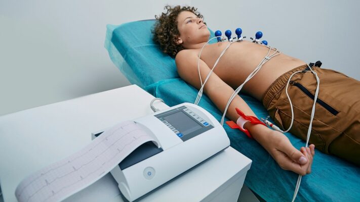
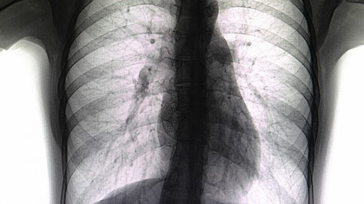
In addition, ECG (Electrocardiography) is used to evaluate the heart’s electrical system. ECG guides us in detecting rhythm disorders. A chest X-ray (Telecardiography) helps us assess the size of the heart within the chest cage and the blood flow status of the lungs. Also, the “Pulse Oximeter” screening that should be performed in every baby in the newborn period is life-saving in the early detection of critical heart diseases that are not yet externally apparent but present with low blood oxygen levels.
The basic tools used in the diagnostic process are:
- Echocardiography
- Electrocardiography
- Telecardiography
- Pulse Oximeter
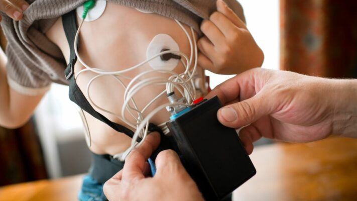
- Holter Monitoring (24-hour rhythm recorder)
- Exercise Test
- In more complex heart diseases
- Transesophageal Echocardiography (TEE)
- Computed Tomography (CT)
- Magnetic Resonance Imaging (MRI)
- Angiography
Do Heart Holes (VSD, ASD) Close on Their Own?
Conditions known among the public as “a hole in the heart” are actually among the most common situations we encounter medically. It is the presence of an opening in the walls that separate the heart’s oxygen-rich and oxygen-poor blood. The location and size of these holes directly affect the course of the condition and the treatment decision.
Ventricular Septal Defect (VSD): This is a hole in the wall between the heart’s lower chambers, the ventricles. It is the most common congenital heart disease. If the hole is small and within the muscle tissue, as the child grows and the heart muscle develops, a very large proportion of these holes close spontaneously. During this process, it is sufficient for the child to be checked only at certain intervals. However, if the hole is large, high-pressure blood passes from the left side of the heart to the right side and then to the lungs. This damages the lung vessels and strains the heart. In these large holes, which have a low likelihood of closing, we support the baby with medication, and if we do not get a response, we proceed with closure by surgery or angiography.
Atrial Septal Defect (ASD): This is a hole between the heart’s upper chambers, the atria. Compared to VSD, it is a more “silent” condition. It usually causes no symptoms in infancy, and the child is quite healthy. Most of the time, it is noticed incidentally due to a murmur. Small ASDs can also close over time. However, large holes that do not close can, if untreated, lead to enlargement of the right side of the heart, rhythm disorders, and permanent damage after the ages of 30–40. Therefore, we usually wait until school age, and if it has not closed, we recommend intervention before starting primary school.
Contact us now to get detailed information about treatments and to create an appointment!
Why Does Bluish Discoloration (Cyanosis) Occur in Babies?
Some heart diseases progress with a noticeable bluish discoloration (cyanosis) in the baby’s skin, lips, and nail beds. Under normal conditions, the oxygen-poor blood on the right side of the heart should go to the lungs and be oxygenated, and the oxygen-rich blood on the left side should be distributed to the body. However, in some structural abnormalities this order is disrupted, and oxygen-poor blood mixes into the systemic circulation without being oxygenated. This is what causes the baby’s color to turn bluish.
The best-known example of this group is Tetralogy of Fallot. In this disease, four different problems are present together in the heart:
- A large VSD,
- Narrowing in the vessel going to the lungs,
- Thickening of the right wall of the heart, and
- Displacement of the origin of the aorta.
In these babies, “cyanotic spells” may be seen especially during crying, straining, or upon waking in the morning. The baby suddenly becomes restless, turns a dark blue-purple color, and has difficulty breathing. In this situation, the most important first aid for families is to stay calm and pull the baby’s knees toward the abdomen (knee-chest position). This maneuver helps the spell pass by improving circulation.
Another critical condition is Transposition of the Great Arteries. In this condition, where the main vessels leaving the heart (the aorta and pulmonary artery) are positioned incorrectly, the body is continuously pumped with oxygen-poor blood, and the lungs are continuously pumped with oxygen-rich blood. The survival of these babies depends on the presence of additional holes in the heart and requires urgent medical intervention immediately after birth.
Cyanotic heart diseases generally do not resolve on their own, and surgical repair is essential.
Some findings seen in cyanotic heart diseases include:
- Bluish discoloration of the lips and inside the mouth (tongue)
- Color change in the nail beds
- Increased bluish discoloration during crying
- Clubbing of the fingertips
- Growth retardation
- Easy fatigue
- Urge to squat
Is Open Surgery Necessary for All Heart Diseases?
Thanks to advances in medical technology, it is no longer necessary to open the chest for every heart hole or narrowing. With methods we call interventional cardiology, similar to angiography procedures in adults, it is possible to reach the heart by entering through the vein or artery in the groin.
Especially in closing holes such as ASD and PDA, in some types of VSD, and in opening narrowed heart valves, these methods are often our first choice. During the procedure, special devices resembling an “umbrella” or a “plug” are used to close the holes. In valve stenosis, a deflated balloon is advanced through the vessel and inflated right at the valve to relieve the narrowing.
The biggest advantage of these methods is that there is no incision scar on the child’s chest. Also, the recovery process is incredibly fast; the child is usually discharged one day after the procedure and can return to normal life and school within a few days. However, not every patient may be suitable for this method. Criteria such as whether the edges of the hole have enough tissue to hold the device or whether the vascular structure is suitable are evaluated. If the anatomy is not suitable, the surgical method becomes the safest option.
How Do Surgical Treatment and the Follow-up Process Work?
Heart surgery (operation) is required for large holes that cannot be closed by angiography, complex cyanotic diseases, or valve replacements. Although the idea of “open-heart surgery” is very frightening for families, success rates in pediatric cardiac surgery centers today are extremely high. Surgeons use very delicate techniques appropriate to the size of the baby’s heart.
The timing of surgery is of vital importance. Some babies are operated on immediately after birth, some at a few months old, and some in the preschool period. The aim is to resolve the problem before permanent organ damage (especially in the lungs) occurs and before the child’s psychology is affected. After a successful surgery, once the intensive care and ward process is completed, children generally reach an exercise capacity similar to their peers.
What Should Be Considered in Home Care for a Child with Heart Disease?
After your child is diagnosed—whether you are in the follow-up period or in the post-operative recovery period—there are some “golden rules” you should pay attention to at home. These rules are to improve your child’s quality of life and protect them from possible complications.
Nutrition:
Babies with heart disease, especially those with heart failure, expend tremendous energy even just to breathe and keep their hearts working. Therefore, they may be weaker than their peers. To close this gap, they need high-calorie nutrition programs. Breast milk is always the priority, but when it is not enough, do not hesitate to use the calorie-enriched special formulas or supplements your doctor recommends.
Oral and Dental Health:
This issue is often overlooked, but it is of vital importance. Decayed teeth or unhealthy gums are an entry point for bacteria to enter the bloodstream. These bacteria can travel through the blood and adhere to the damaged area in the heart, the edge of a hole, or an artificial valve/patch if present, causing a very serious heart infection called “Infective Endocarditis.” Therefore, in children with heart disease, brushing teeth is at least as important as medications.
Important dental care recommendations are:
- Brushing twice a day
- Regular dental check-ups
- Treating cavities without delay
- Using dental floss
Protection from Infections:
The lungs of children with heart disease are more sensitive, and even an ordinary flu can turn into severe pneumonia in them. Therefore, it is essential that childhood vaccinations are completed in full. In addition, an annual flu vaccine is recommended for every child with heart disease older than 6 months. Beyond that, for babies born prematurely or with severe heart failure, monthly protective injections (immunization program) against the RSV virus are administered in the winter season. This is a great opportunity to get through the winter months without being hospitalized.
Physical Activity:
The biggest fear of families is the question, “If my child runs, will something happen to their heart?” There is usually no harm in children who have had surgery and completely recovered or who have a small hole doing sports; on the contrary, activity is necessary for heart health. Only in children with severe stenosis, rhythm disorders, or heart muscle disease may we restrict competitive sports. However, the child is generally allowed to play at their own pace and ride a bicycle. The most accurate information on this will be given by your doctor, who knows your child’s exercise test and ECHO results.
Frequently Asked Questions
What causes congenital heart diseases in children?
Congenital heart diseases in children are caused by structural abnormalities that occur during the development of the heart in the womb. Genetic predisposition, chromosomal anomalies, and family history can increase the risk. In some cases, environmental factors also contribute to the process.
Do infections during pregnancy increase the risk of a hole in the heart in children?
Some viral infections such as rubella during pregnancy, uncontrolled diabetes, and the use of harmful medications can affect the baby’s heart development. This can increase the risk of a hole in the heart or valve anomalies, and regular prenatal follow-up is important.
Which symptoms in children with a hole in the heart, and when do they appear?
If the hole in the heart is small, it may not cause symptoms for a long time. In large holes, rapid breathing, bluish discoloration, failure to gain weight, and frequent infections may be seen. Symptoms are usually noticed in infancy, but some conditions may appear at school age.
How are growth and development affected in children with valve stenosis?
Valve stenosis increases the workload of the heart to pump blood, which can cause the child to tire easily and fail to gain adequate weight. Growth retardation may be seen in long-term and severe stenosis; therefore, regular cardiology follow-up is required.
Is it safe for children with congenital heart disease to do sports?
Sports restrictions vary depending on the type and severity of the disease. While controlled exercise may be recommended in mild cases, strenuous exertion should be avoided in children with severe valve stenosis or rhythm problems. The decision should be made by a pediatric cardiologist.
What is the success rate in congenital heart diseases that require surgery?
Today, success rates in pediatric cardiac surgery are quite high. With early diagnosis and interventions performed at an appropriate center, most children can reach a normal life expectancy. However, long-term follow-up is important in complex cases.
How long does the recovery process take in children after heart surgery?
The length of hospital stay after surgery usually varies from a few days to a few weeks. Full recovery may take a few months. During this period, protection from infections, nutrition, and doctor check-ups play a critical role.
Can congenital heart disease lead to other heart problems in the future?
In untreated or severe diseases, arrhythmias, heart failure, and increased pulmonary pressure may develop. Even after successful treatment, some children may require regular cardiology follow-up in adulthood.
What complications can valve regurgitation cause in children?
Valve regurgitation can lead to enlargement of the heart over time and a decrease in pumping strength. In advanced cases, shortness of breath, easy fatigue, and rhythm problems may occur. Early intervention reduces the risk of complications.
How should the psychological development of children with congenital heart disease be supported?
Frequent hospital visits and physical restrictions can cause anxiety and self-confidence issues in the child. Family support, open communication, and, when needed, psychological counseling positively affect the child’s social adaptation and quality of life.
Blog Posts

Exercise Stress Test in Children: Purpose, Procedure and Results
Exercise stress testing in children is [...]
Read More
29
Sep
Sep
Holter ECG Monitoring in Children: Procedure, Uses and Results
Holter ECG in children is a [...]
Read More
29
Sep
Sep

Pediatric Echocardiography (ECHO): Procedure, Uses and Results
Pediatric echocardiography is a painless ultrasound [...]
Read More
29
Sep
Sep
Pediatric ECG (Electrocardiogram): Procedure, Uses and Results
Pediatric ECG (Electrocardiography) is a completely [...]
Read More
29
Sep
Sep

Pulmonary Valve Stenosis: Symptoms, Causes, Diagnosis and Treatment
Pulmonary valve stenosis (pulmonary stenosis) is [...]
Read More
29
Sep
Sep

Ventricular Septal Defect (VSD): Symptoms, Causes and Treatment Options
Ventricular septal defect (VSD) is a [...]
Read More
29
Sep
Sep
Atrial Septal Defect (ASD): Symptoms, Causes and Treatment Options
Atrial septal defect (ASD) is a [...]
Read More
29
Sep
Sep

Shortness of Breath in Children: Causes, Symptoms and Treatment Options
Shortness of breath in children is [...]
Read More
10
Aug
Aug