
 Türkçe
 Deutsch
 العربية
 Русский

Acyanotic heart diseases in children (non-cyanotic heart diseases) are congenital heart defects in which, despite the presence of a structural abnormality in the heart, the oxygen level in the blood does not decrease significantly. In these diseases, cyanosis is not observed; however, the workload of the heart increases and the circulatory system is affected.
Septal defects such as ventricular septal defect (VSD) and atrial septal defect (ASD) (holes in the heart) are among the most common causes of acyanotic heart diseases. In this condition, oxygenated blood on the left side of the heart passes to the right side, leading to excessive blood flow to the lungs and, over time, signs of heart failure may develop.
Congenital heart diseases such as patent ductus arteriosus (PDA) and atrioventricular septal defect are among the other most common acyanotic heart diseases and cause volume overload in the pulmonary circulation. Especially during infancy, they may present with clinical symptoms such as frequent respiratory infections, feeding difficulties, and poor weight gain.
In acyanotic heart diseases, diagnosis is confirmed by echocardiography after a heart murmur is detected during physical examination. Without early diagnosis and appropriate medical or surgical treatment, unfortunately, the risk of developing pulmonary hypertension and permanent cardiac damage increases.
| What You Need to Know | Information |
| Definition | Acyanotic heart diseases in children are congenital heart diseases that lead to abnormal direction of blood flow or cardiac overload due to congenital anomalies in the heart chambers or major vessels, but without a significant mixing of deoxygenated blood into the systemic circulation. |
| Basic Pathophysiology | Usually there is a left-to-right shunt (oxygenated blood redirected back to the lungs) or obstructive lesions (narrowing in blood flow). Pulmonary hypertension and heart failure may develop in the long term. |
| Main Types | Atrial septal defect (ASD), ventricular septal defect (VSD), patent ductus arteriosus (PDA), atrioventricular septal defect (AVSD), pulmonary valve stenosis, peripheral pulmonary stenosis, aortic stenosis, aortic coarctation |
| Frequency | They constitute a significant portion of congenital heart diseases. The most common form is ventricular septal defect. |
| Risk Factors | Genetic syndromes (e.g., Down syndrome), family history, maternal diabetes, infections during pregnancy (e.g., rubella), exposure to teratogenic medications. |
| Clinical Findings | Rapid breathing, feeding difficulty, poor weight gain, sweating, easy fatigue, frequent lung infections, heart murmur. Cyanosis is usually not observed. |
| Physical Examination Findings | Heart murmur, tachycardia, hepatomegaly (if heart failure has developed), differences in arm and leg pulses (especially in aortic coarctation), growth and developmental delay. |
| Diagnostic Methods | Echocardiography (primary diagnostic tool), electrocardiography (ECG), chest X-ray, pulse oximetry, and when necessary cardiac MRI or cardiac CT and angiography (catheterization). |
| Hemodynamic Effect | In diseases with left-to-right shunt, pulmonary blood flow increases; in obstructive lesions, pressure overload develops in the affected chamber. |
| Complications | Pulmonary hypertension, Eisenmenger syndrome (cyanosis may develop in advanced stages), heart failure, growth and developmental delay, arrhythmia. |
| Treatment Approaches | Follow-up (spontaneous closure may occur in small defects), mild stenosis may resolve spontaneously, medical therapy (diuretics, ACE inhibitors), catheter interventions, surgical correction. |
| Surgical Indication | Determined according to the size of the defect, the patient’s age, symptoms, pulmonary artery pressure, and signs of heart failure. |
| Prognosis | With early diagnosis and appropriate treatment, life expectancy and quality of life are quite good in most patients. In untreated cases, the risk of long-term complications increases. |
| Follow-Up Requirement | Regular pediatric cardiology follow-up is necessary. Long-term monitoring after intervention is recommended. |
| Preventive Measures | Pre-pregnancy counseling, control of maternal diseases, protection from infections, and regular prenatal follow-up are important. |

PROF. DR.
İbrahim Cansaran TANIDIR
Pediatric Cardiology Specialist
İbrahim Cansaran TANIDIR
Pediatric Cardiology Specialist
Prof. Dr. İbrahim Cansaran Tanıdır is an internationally recognized expert with nearly 20 years of clinical, academic, and interventional experience in congenital heart diseases, pediatric interventional cardiology, hybrid procedures, and radiation-reduced / fluoroscopy-free catheterization techniques.
To date, he has performed more than 10,000 pediatric cardiac catheterization procedures both in our country and abroad; and has pioneered advanced interventional and hybrid applications in complex structural heart diseases in our country. His academic productivity represents a strong scientific authority with 110 SCI/SCIE publications, 1,440 citations, and an H-index of 18 (Google Scholar, 2025).
Why Do Acyanotic Heart Diseases Not Cause Cyanosis?
To understand this, we need to visualize the traffic inside the heart. Normally, the heart sends deoxygenated blood from the right side to the lungs, while oxygenated blood is pumped to the body from the left side. There should be no transition between the two sides. In acyanotic heart diseases, however, there is usually a leakage from the high-pressure region on the left side of the heart (where oxygenated blood is located) to the low-pressure region on the right side (where deoxygenated blood is located).
In medical terms, we call this a “left-to-right shunt.” In other words, some of that valuable, oxygen-rich red blood that should go to the body takes the wrong path and returns to the lungs again. Since the blood is already oxygenated and re-enters the circulation without becoming deoxygenated, we do not observe bluish discoloration or darkening in the baby’s color. The blood remains red.
However, the absence of cyanosis does not mean everything is normal. This condition causes excessive blood flow to the lungs. The lung vessels have difficulty carrying this excessive load, and over time an increase in pressure occurs. Another scenario is when there is no leakage but narrowing in the pathways through which blood passes (valves or vessels). In this case the blood is still oxygenated, but the heart must exert much more force to pump it. As a result, in acyanotic diseases the main problem is not lack of oxygen but the extra workload placed on the heart and lungs.
Which Symptoms at Home May Indicate Acyanotic Heart Diseases?
Since there is no obvious “alarm” color such as cyanosis in the baby, the symptoms of acyanotic heart diseases are usually hidden in the baby’s general condition and exercise tolerance. In the newborn period, lung resistance is high, so babies may show no symptoms during the first days. However, when the baby is a few weeks old, the lung vessels relax and the amount of blood passing through the hole in the heart increases. That is when some subtle signs begin to appear.
The most common signs that attentive parents may observe include:
- Rapid breathing
- Becoming tired quickly while feeding or breastfeeding
- Bead-like sweating on the forehead during feeding
- Frequent need to pause and rest while feeding
- Stagnation in weight gain
- Frequent recurrent lung infections
- Wheezing breathing
- Reluctance to feed
- Fatigue
- Mild swelling of the eyelids
- Symptoms such as abdominal pain or colic
- Paleness
These symptoms are actually the baby’s way of saying “I am getting tired.” The heart works so hard to pump blood to the body that the baby may not even tolerate the energy required just for feeding. If several of these symptoms are present together in your baby, it is important not to dismiss them as simple loss of appetite or colic but to have the heart evaluated.
Is a Murmur Heard During Examination Always an Acyanotic Heart Disease?
One of the sentences you hear most often during a doctor’s visit and that causes the most concern is “I hear a murmur in the heart.” However, there is no need to panic immediately. A murmur is not actually the name of a disease; it is simply a sound we hear while listening to the heart. It refers to the sound produced by blood flowing through the heart.
In many children, even when there is no hole or narrowing in the heart, sounds known as “innocent murmurs” may be heard. Children have thin chest walls, their hearts beat rapidly, and blood flow is dynamic; this can create vibrations that are sometimes heard with a stethoscope. Innocent murmurs are generally not a sign of disease.
However, murmurs heard in acyanotic heart diseases are different. These sounds result from turbulence created when blood passes under pressure through a hole (such as VSD or PDA) or struggles to pass through a narrowed valve. Pathological murmurs are usually louder, the tone of the sound is harsher, and they may sometimes radiate to the back or neck. Your doctor can largely distinguish whether the murmur is innocent or indicates a problem by evaluating its character, timing, and intensity. The definitive distinction is of course made with echocardiography.
How Does VSD, the Most Common Acyanotic Heart Disease, Progress?
Ventricular Septal Defect, or VSD for short, is the most common condition among all congenital heart diseases. It is popularly known as a “hole between the ventricles of the heart.” It is an opening in the wall between the lower two chambers of the heart.
The course of VSD depends entirely on the size and location of the hole. If the hole is small, the child usually has no symptoms. The child grows, runs, and plays normally. Only the doctor hears a very loud murmur during examination. Interestingly, the smaller the hole, the louder the sound produced when blood passes through it. Most of these small VSDs, especially those located in the muscular tissue, close spontaneously as the child grows. For this reason, we do not immediately recommend surgery but follow the child patiently.
However, if the hole is large, the situation is different. In large VSDs, the amount of blood passing from the left side to the right side and then to the lungs is very high. In these babies, signs of heart failure begin early, usually around 1–2 months of age. The baby cannot gain weight and experiences shortness of breath. In such cases we first try to relieve the heart with medications. If the baby cannot grow despite medication or if lung pressure increases, the hole must be closed either surgically or, if appropriate, through an angiographic method. In short, not every child diagnosed with VSD undergoes surgery or angiography; most grow up with regular follow-up alone.
Contact us now to get detailed information about treatments and to create an appointment!
Why Is ASD in the Acyanotic Group a Disease That Progresses Silently?
Atrial Septal Defect (ASD) is a hole between the upper chambers of the heart, called the atria. There is a very valid reason why we call this disease “silent”: it almost never produces symptoms during childhood. Unlike babies with VSD, children with ASD are generally very comfortable. Their weight gain is normal, they do not experience shortness of breath, and they can run and play. Families are often surprised and say, “My child had no problems at all, how can there be a hole in the heart?”
This “silence” is somewhat dangerous because it may delay diagnosis. In ASD, the pressure difference between the atria is low, so the murmur may be very faint or not heard at all. Suspicion usually arises when a careful physician notices a fixed splitting of the second heart sound (a heart examination finding).
If it is not detected in childhood, permanent damage such as enlargement of the right side of the heart, rhythm disorders, and increased lung pressure may begin to appear in the 30s or 40s. Therefore, when we diagnose ASD, even if the child has no symptoms, if there is enlargement on the right side of the heart we usually recommend closing it during the preschool period (around 4–5 years of age). Today this procedure can often be performed without surgery by entering through the groin using devices called “umbrellas.”
Is PDA Seen in Premature Infants an Acyanotic Heart Disease?
Yes. Patent Ductus Arteriosus (PDA) is also included in this group. The ductus is a vital vessel while the baby is in the womb; it forms a bridge between the aorta and the pulmonary artery. When the baby is born and takes the first breath, the role of this vessel ends and it should close. However, especially in premature babies, it may remain open because the vessel structure has not matured.
This opening causes oxygenated blood to continuously leak from the aorta into the pulmonary artery. The most typical finding of PDA is a continuous murmur that resembles the sound of a “machine.” In addition, these babies have very bounding pulses, a condition we call “water-hammer pulse.”
In premature infants, our first line of treatment for PDA is medication. With drugs such as ibuprofen or paracetamol we can cause this vessel to constrict and close. However, in full-term babies medications usually do not work.
If the opening is large enough to strain the heart, it must be closed using non-surgical methods, usually by placing a vascular plug. PDA is one of the diseases with the most successful treatment outcomes and complete recovery.
Is Narrowing in the Aorta (Coarctation) Included in the Acyanotic Group?
Aortic coarctation is also included in the acyanotic group, but its mechanism is different from the others. Here there is not a hole or leakage but an “obstruction.” There is narrowing in a part of the main artery (aorta) leaving the heart.
This narrowing blocks blood flow like a dam. Behind the narrowing (in the head and arms) pressure rises significantly, while beyond the narrowing (in the abdomen and legs) pressure and blood flow decrease. This disease is very critical for us because in newborn babies it may create a sudden crisis when the ductus closes. The baby may go into shock, urine output may decrease, and severe metabolic acidosis may develop.
The most important clue during examination is checking the baby’s leg pulses (femoral pulses). If pulses are strong in the arms but absent or weak in the groin, aortic coarctation is suspected.
In older children it may be detected incidentally while investigating hypertension (high blood pressure). Depending on the location and type of narrowing and the age and weight of the patient, balloon angioplasty, stent placement, or surgical methods are used in treatment.
Which Tests Are Requested for the Diagnosis of Acyanotic Heart Diseases?
Physical examination and listening with a stethoscope tell us a lot, but we rely on technology for a definitive diagnosis. We need to see inside the heart, measure flow velocities, and determine the exact location of the defect.
The main tests your doctor may request include:
- Echocardiography (ECHO): The most important diagnostic tool. It visualizes the heart using ultrasound waves. It does not contain radiation and is painless.
- Electrocardiography (ECG): Shows the electrical system and rhythm of the heart. It helps us understand thickening of the heart walls (hypertrophy). It does not contain radiation and is painless.
- Chest X-ray: Shows the size of the heart shadow and the intensity of blood flow to the lungs.
- Cardiac MRI or CT: May rarely be required to visualize more complex anatomical structures.
- Cardiac Catheterization: Usually performed not only for diagnosis but also for treatment (closing the defect or relieving narrowing).
None of these tests (except catheterization) are painful or invasive. Families should not worry about how these tests will be performed on a tiny baby; the procedures are usually completed while the baby lies calmly on a bed or in the mother’s arms.
How Should a Baby with Acyanotic Heart Disease Be Fed?
In babies with heart disease, our biggest challenge is nutrition. Imagine a baby with a large VSD or PDA; the heart works so fast and the baby makes so much effort to breathe that the metabolism functions 40–50% faster than peers. In other words, your baby is actually running a marathon continuously while lying still.
To meet this high energy requirement, they need much more calories than normal. However, the problem is that breastfeeding also requires effort, and these babies get tired very quickly while feeding. Because they tire quickly, they fall asleep before their stomach is full. This vicious cycle prevents weight gain.
Important points in feeding these babies include:
- Small and Frequent Feeding: Meals should be divided so the stomach does not become too full and put pressure on the diaphragm.
- Calorie Enrichment: Adding special energy powders to breast milk or formula, or using high-calorie formulas directly.
- MCT Oil: Increasing calorie density by adding special fats that are easy to digest.
- Keeping Breastfeeding Duration Short: Avoid tiring the baby by keeping them on the breast longer than 20–30 minutes.
- Nasogastric Tube: If the baby becomes very tired and loses weight, feeding through a thin tube inserted from the nose to the stomach.
When a nasogastric tube is recommended, families often feel very upset and see it as a setback. However, it is actually a temporary and very beneficial support method that allows the baby to save energy for growth instead of spending it on feeding. As the baby becomes stronger and gains weight, the tube is removed and normal feeding resumes. Remember, having a good weight on the day of surgery will also speed up the recovery process.
Contact us now to get detailed information about treatments and to create an appointment!
How Do Infections Affect the Course of Acyanotic Heart Diseases?
In acyanotic heart diseases, especially in large defects, the blood flow going to the lungs is increased. Lung vessels are fuller than normal and the tissues are more “wet” (edematous). Unfortunately, this moist and highly perfused environment becomes an excellent breeding ground for viruses and bacteria.
In a healthy child, a simple flu infection that passes with only a runny nose may rapidly turn into pneumonia or trigger heart failure in a child with heart disease. Therefore, protection from infections is vitally important for us.
To manage the risk of infection, the following should be done:
- Hand Washing: Everyone should wash their hands before touching the baby.
- Avoiding Crowds: Especially during winter months, crowded indoor places such as shopping malls should be avoided.
- RSV Prophylaxis (Synagis): Protective antibody injections administered monthly during the winter season (October–March).
- Routine Vaccinations: The vaccination schedule recommended by the Ministry of Health should be followed without interruption.
- Influenza Vaccine: Annual influenza vaccination for children with heart disease older than 6 months and their caregivers.
- Dental Health: Bacteria from decayed teeth can enter the bloodstream and settle in the heart (Endocarditis). Therefore tooth brushing and dental check-ups are very important.
In the past, antibiotics were given to every heart patient before dental extraction or filling. According to the current 2024–2025 guidelines, this practice is now only applied to very high-risk patients (those who previously had endocarditis, those with prosthetic valves, etc.). However, the best protection is not antibiotics but good oral hygiene.
Is Surgery Always Necessary in the Treatment of Acyanotic Heart Diseases?
The first question every family asks after receiving a diagnosis of acyanotic heart disease is “Will surgery be required?” The answer is not always yes. The treatment strategy is determined entirely according to the type and severity of the disease and the condition of the child.
Small VSDs, mild stenosis (such as pulmonary stenosis), or small PDAs usually require no intervention. These children are simply called for follow-up at certain intervals. Often the defects close spontaneously or the narrowing becomes insignificant as the child grows.
In cases where intervention is necessary, modern medicine offers highly advanced options. With methods known as “Interventional Cardiology,” many procedures can be performed through angiography via the groin without opening the chest. For example, suitable VSDs, ASDs, and PDAs can be closed within minutes using devices similar to an “umbrella” or “plug.” In valve stenosis, balloon dilation (valvuloplasty) or stent placement may be performed.
Surgery (open heart surgery) is generally preferred if the defect is very large, unsuitable for catheter closure, or accompanied by other problems. The goal of surgery is complete correction. In other words, the anatomy of the heart is restored to normal rather than simply patched. Surgical success rates today are around 98–99%. Although the decision for surgery may seem frightening, it is actually the key that opens the door for the child to live a lifetime with a healthy heart.
Can a Child with Acyanotic Heart Disease Do Sports After Recovery?
This is perhaps the part that affects children’s psychology the most. With a natural instinct to protect, families tend to restrict their children by saying “don’t run” or “don’t get tired.” This can leave the child physically weaker and cause them to be labeled as the “sick child” among peers.
The general rule is this: patients with acyanotic heart disease who have been successfully treated (whether closed with a device or surgically) and who no longer have leakage or pressure overload in the heart can perform all kinds of sports just like normal children. They can become professional football players, swim, run. Once the heart muscle has recovered, it is no different from a normal heart.
However, there are some special situations and considerations regarding sports:
- Completely Corrected Patients: Usually no restrictions.
- Mild Stenosis: No restriction is required in isolated mild valve stenosis.
- Severe Aortic Stenosis/Coarctation: Even if treated, “static exercises” such as weightlifting, wrestling, or heavy lifting that create sudden and high pressure may be prohibited.
- Patients Using Blood Thinners: Sports with a risk of trauma such as boxing or karate should be avoided.
Consult your doctor about the sports your child can participate in according to their condition.
Frequently Asked Questions
Which structural abnormalities cause acyanotic heart diseases in children?
Acyanotic heart diseases in children usually develop due to holes in the heart wall, valve stenosis, or abnormal vascular connections. These conditions lead to mixing of oxygenated and deoxygenated blood or cause the heart to work under excessive load.
Which early symptoms reveal acyanotic heart diseases in infants?
Symptoms such as rapid breathing, quick fatigue during feeding, excessive sweating, poor weight gain, and frequent lung infections may be observed in infants. The absence of cyanosis may delay diagnosis, so growth retardation is an important clue.
Which risk factors are associated with acyanotic heart diseases in children?
Genetic predisposition, consanguineous marriage, maternal infections during pregnancy, diabetes, or medication use are among the risk factors. They may also occur more frequently with certain chromosomal disorders such as Down syndrome.
How do acyanotic heart diseases in children affect lung pressure?
In these diseases, the high pressure on the left side of the heart may be transmitted to the right side and the lungs. If left untreated for a long time, pulmonary vascular pressure may increase and permanent pulmonary hypertension may develop.
What complications may develop if acyanotic heart diseases in children progress?
If untreated, heart failure, rhythm disorders, and growth retardation may occur. In advanced cases, lung pressure may increase permanently and the possibility of surgery may decrease.
Which advanced examinations are used in the diagnosis of acyanotic heart diseases in children?
Echocardiography is the primary diagnostic method. When necessary, cardiac catheterization, MRI, or CT angiography may be performed. These tests enable detailed evaluation of the heart structure and pressure values.
In which situations do acyanotic heart diseases in children require surgery?
Large heart defects, severe valve stenosis, or vascular anomalies that strain the heart may require surgery. The decision for surgery is made by evaluating the child’s age, weight, and the burden on the heart.
What is the recovery process like after surgery for acyanotic heart diseases in children?
Recovery after surgery is usually completed within a few weeks. Most children can return to normal activities. Regular cardiology follow-ups are important for monitoring heart function.
In which situations can acyanotic heart diseases in children be treated with angiography?
The possibility of treatment with angiography should be evaluated in most children who require surgery. The indications are the same as for surgery. However, not every patient is suitable for angiographic treatment.
What is the recovery process like after angiography in acyanotic heart diseases in children?
Patients are usually discharged one day after the procedure. Most children can return to normal activities within weeks. Regular cardiology follow-ups are important for monitoring heart function.
How can acyanotic heart diseases in children affect school-age life?
In untreated cases, easy fatigue and attention difficulties may occur. Children who receive timely treatment can usually show physical and academic performance similar to their peers.
Do acyanotic heart diseases in children require lifelong follow-up?
Many patients can live healthy lives after successful treatment; however, the treated areas should be monitored regularly. Cardiology follow-up may also be recommended in adulthood.
Blog Posts
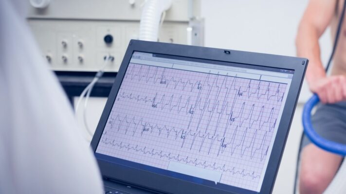
Exercise Stress Test in Children: Purpose, Procedure and Results
Exercise stress testing in children is [...]
Read More
29
Sep
Sep
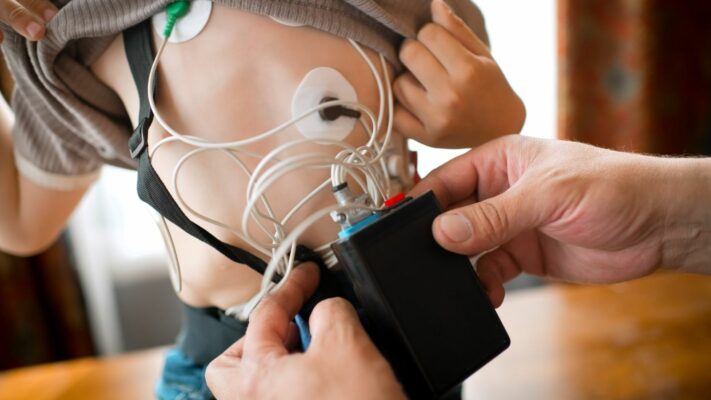
Holter ECG Monitoring in Children: Procedure, Uses and Results
Holter ECG in children is a [...]
Read More
29
Sep
Sep
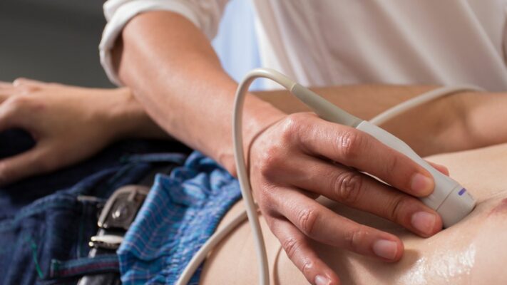
Pediatric Echocardiography (ECHO): Procedure, Uses and Results
Pediatric echocardiography is a painless ultrasound [...]
Read More
29
Sep
Sep
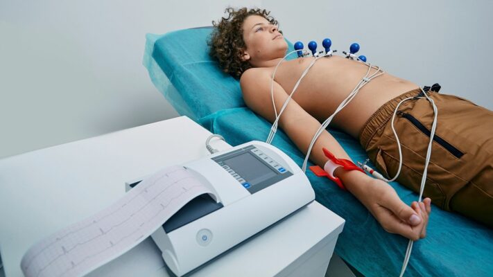
Pediatric ECG (Electrocardiogram): Procedure, Uses and Results
Pediatric ECG (Electrocardiography) is a completely [...]
Read More
29
Sep
Sep

Pulmonary Valve Stenosis: Symptoms, Causes, Diagnosis and Treatment
Pulmonary valve stenosis (pulmonary stenosis) is [...]
Read More
29
Sep
Sep
Ventricular Septal Defect (VSD): Symptoms, Causes and Treatment Options
Ventricular septal defect (VSD) is a [...]
Read More
29
Sep
Sep
Atrial Septal Defect (ASD): Symptoms, Causes and Treatment Options
Atrial septal defect (ASD) is a [...]
Read More
29
Sep
Sep

Shortness of Breath in Children: Causes, Symptoms and Treatment Options
Shortness of breath in children is [...]
Read More
10
Aug
Aug