
 Türkçe
 Deutsch
 العربية
 Русский

Heart diseases in children accompanied by cyanosis are characterized by bluish discoloration of the lips, nails, and skin due to decreased oxygen levels in the blood. This condition is most often caused by congenital heart anomalies and can lead to serious respiratory and circulatory problems if not diagnosed early.
Among congenital cyanotic heart diseases, Tetralogy of Fallot, transposition of the great arteries, and tricuspid atresia are commonly observed. In these diseases, due to the mixing of deoxygenated and oxygenated blood, the oxygen content in the blood decreases and sufficient oxygen cannot reach the systemic circulation. The clinical picture may become evident with cyanotic attacks and growth retardation.
In infants, signs of cyanosis may increase especially during crying, feeding, or exertion. Findings accompanying cyanosis such as rapid breathing, frequent lung infections, easy fatigue, and poor weight gain should be carefully evaluated. Physical examination and oxygen saturation measurement constitute the basic steps in the diagnostic process.
Treatment of cyanosis in children and surgical/angiographic approaches are planned according to the type and severity of the disease. Medication therapy, oxygen support, and corrective heart surgeries are the main options. Early surgical intervention is the most important factor that significantly increases life expectancy and quality of life.
| What You Need to Know | Information |
| Definition | Cyanosis is a clinical finding characterized by bluish discoloration of the skin and mucous membranes (especially inside the mouth/lips) due to decreased oxygen saturation in the blood. In childhood, the most common cause is congenital heart disease. |
| Mechanism of Cyanosis | It usually occurs when low-oxygen blood passes into the systemic circulation as a result of heart anomalies that create a right-to-left shunt. Decrease in arterial oxygen saturation is the main pathophysiological mechanism. |
| Main Congenital Heart Diseases with Cyanosis | Tetralogy of Fallot, Transposition of the Great Arteries (TGA), Tricuspid Atresia, Pulmonary Atresia, Total Anomalous Pulmonary Venous Return (TAPVD), Single Ventricle type diseases (such as Hypoplastic Left Heart Syndrome, Hypoplastic Right Heart Syndrome, Double Inlet Ventricle, Double Outlet Ventricle) |
| Tetralogy of Fallot | It consists of ventricular septal defect (VSD), pulmonary stenosis, dextroposition of the aorta, and right ventricular hypertrophy. Cyanosis usually becomes evident during infancy and hypoxic attacks known as “tet spells” may occur. |
| Transposition of the Great Arteries (TGA) | The aorta arises from the right ventricle, and the pulmonary artery arises from the left ventricle. Oxygenated and deoxygenated blood circulations run in parallel. Mixing at the atrial, ventricular, or ductal level is necessary for survival. |
| Tricuspid Atresia | Due to the absence of development of the tricuspid valve, blood flow to the right ventricle is prevented. Cyanosis develops with a right-to-left shunt. It is usually accompanied by VSD and atrial septal defect (ASD). |
| Pulmonary Atresia | Because the pulmonary valve is completely closed, there is no blood flow from the right ventricle to the lungs. Pulmonary circulation is usually maintained through the ductus arteriosus. |
| Total Anomalous Pulmonary Venous Return (TAPVD) | Pulmonary veins open not into the left atrium but into the systemic venous circulation and from there into the right atrium. A shunt at the atrial level is required for oxygenated blood to enter the systemic circulation. |
| Hypoplastic Left Heart Syndrome | It is characterized by severe underdevelopment of the left heart structures. Systemic circulation is usually dependent on the ductus arteriosus. Severe cyanosis and circulatory disorder occur in the neonatal period. |
| Clinical Findings | Bluish discoloration of the lips, tongue, and nail beds; feeding difficulty; rapid breathing; easy fatigue; growth retardation; fainting attacks. |
| Tet Attacks (Hypoxic Spell) | These are sudden hypoxia attacks especially seen in Tetralogy of Fallot. They progress with restlessness, deep cyanosis, and changes in consciousness. Emergency intervention is required. |
| Diagnostic Methods | Physical examination, measurement of oxygen saturation with pulse oximetry, electrocardiography (ECG), chest X-ray, echocardiography (ECHO), and when necessary cardiac catheterization and advanced imaging methods. |
| Importance in the Neonatal Period | Severe cyanotic heart diseases that are not noticed early can be life-threatening. Oxygen saturation measurement in newborn screening is of critical importance. |
| Treatment Approach | Treatment varies according to the disease. Keeping the ductus open with Prostaglandin E1, placing a stent in the ductus, surgical corrective operations, palliative interventions, and in some cases heart transplantation may be required. Many repair or palliation options are available depending on the type of disease. |
| Surgical Timing | It may be planned in the neonatal period or in the first months depending on the type and severity of the disease. In some diseases, staged surgical procedures are applied. |
| Long-Term Follow-Up | Regular cardiology check-ups, monitoring of growth and development, evaluation of exercise capacity, and follow-up of possible complications are required. |
| Possible Complications | Heart failure, rhythm disorders, brain abscess, stroke, polycythemia, growth retardation. |
| Prognosis | With early diagnosis and appropriate surgical intervention, life expectancy and quality of life have significantly increased in many cyanotic heart diseases. However, prognosis depends on the type of disease and accompanying anomalies. |

PROF. DR.
İbrahim Cansaran TANIDIR
Pediatric Cardiology Specialist
İbrahim Cansaran TANIDIR
Pediatric Cardiology Specialist
Prof. Dr. İbrahim Cansaran Tanıdır is an internationally recognized expert with nearly 20 years of clinical, academic, and interventional experience in congenital heart diseases, pediatric interventional cardiology, hybrid procedures, and radiation-reduced / fluoroscopy-free catheterization techniques.
To date, he has performed more than 10,000 pediatric cardiac catheterization procedures both in our country and abroad; and has pioneered advanced interventional and hybrid applications in complex structural heart diseases in our country. His academic productivity represents a strong scientific authority with 110 SCI/SCIE publications, 1,440 citations, and an H-index of 18 (Google Scholar, 2025).
What Causes the Bluish Discoloration on My Child’s Skin?
First, we need to understand the mechanism of cyanosis, in other words, how it works behind the scenes. In our blood, there are special transport vehicles called “hemoglobin” responsible for carrying oxygen. When these vehicles load oxygen from the lungs, the color of the blood turns into a bright, vivid red. This gives your child’s skin and lips that healthy pink color. However, when these transport vehicles unload their cargo, meaning oxygen, to the tissues, or when they start their journey without receiving oxygen from the lungs, their color darkens and turns bluish-purple.
For physicians, the visibility of cyanosis depends on a mathematical threshold. When the amount of deoxygenated hemoglobin in the blood exceeds a certain level (usually 5 grams per deciliter), we begin to notice the bluish discoloration with the naked eye. This condition generally occurs when the oxygen saturation in the blood falls below 90% or 85%. However, there is a very important risk of misinterpretation here that you should definitely know.
If your child has anemia, meaning the total number of transport vehicles in the blood is already low, cyanosis may not appear even if the oxygen level drops to dangerously low levels (even down to 60%). This is because there is not enough hemoglobin to turn bluish. We call this a “pale danger”; the child may only appear pale but is actually suffering from severe oxygen deprivation. Conversely, in newborn babies with very high blood counts (polycythemia), even a slight drop in oxygen level can make the baby appear intensely purple. In other words, not every cyanosis indicates the same urgency, and the absence of cyanosis does not necessarily mean everything is normal.
Is Every Bluish Discoloration a Sign of Heart Disease?
This question is one of the first asked by concerned families. Our answer is a clear “no.” When evaluating cyanosis, the first thing we do is determine whether it is “central” or “peripheral.” This distinction is the biggest step we take toward diagnosis.
Peripheral cyanosis usually occurs due to harmless reasons. It appears on the child’s hands and feet while the tongue and inside of the mouth remain pink. When a baby gets cold, cries excessively, or becomes frightened, the body says “I must protect vital organs first” and constricts the vessels in the hands and feet. As blood flow slows, the tissues consume the oxygen in the blood and that area becomes bluish. However, the oxygen level of the blood leaving the heart is actually normal.
The condition that truly alarms us and suggests heart disease is “central cyanosis.” In this case, the bluish discoloration appears not only on the fingertips but also on the tongue, lips, oral mucosa, and sometimes throughout the entire body. A purple tongue is a very strong warning sign for us. It indicates that the blood leaving the heart is already oxygen-poor from the beginning.
The main conditions that can cause central cyanosis include:
- Congenital heart diseases
- Severe lung infections
- Airway obstructions
- Suppression of breathing after brain hemorrhage or trauma
- Rare blood disorders such as methemoglobinemia
How Can Heart-Related and Lung-Related Cyanosis Be Distinguished?
When a newborn baby turns blue, it is vital to understand whether it is due to insufficient ventilation of the lungs (such as pneumonia or transient tachypnea of the newborn) or because the heart mistakenly pumps deoxygenated blood into the body. Sometimes it may be difficult to make this distinction just by looking at the baby, but we have some clues.
Interestingly, babies with heart-related cyanosis may appear “comfortable.” The baby may be deeply blue and have low oxygen levels, yet may not show excessive breathing effort such as chest retractions, grunting, or nasal flaring, which are commonly seen in babies with lung disease. Despite appearing comfortable, the baby’s color is poor.
To make a definite distinction, physicians use a method called the “Hyperoxia Test.” We give the baby 100% pure oxygen through a mask. The logic is this: if the problem is in the lungs, when we provide high-pressure oxygen externally, the lungs will mix it into the blood and the baby’s color will improve, and blood gas values will correct. However, if the problem is a structural defect in the heart or reversed vessels, meaning deoxygenated blood bypasses the lungs and circulates through the body, no matter how much oxygen we provide, the blood cannot meet oxygen because it does not reach the lungs. Therefore, the baby’s color will not improve and oxygen levels will not rise. This result tells us, “The problem is in the heart; a cardiologist must see the baby urgently.”
How Do Cyanotic Heart Diseases and Deoxygenated Blood Mixing Occur?
In a normal circulatory system, the oxygen-depleted “deoxygenated” blood coming from the body reaches the right side of the heart, goes to the lungs to be cleaned, passes to the left side, and is then distributed throughout the body in a completely oxygenated form. In other words, deoxygenated and oxygenated blood never mix. In cyanotic heart diseases, this remarkable order is disrupted.
The main problem is the phenomenon we call a “right-to-left shunt.” Due to holes in the heart or abnormal placement of vessels, deoxygenated blood coming from the body crosses directly to the side where oxygenated blood is present and is pumped into the body without having the chance to be cleaned in the lungs. As a result, the blood circulating in the child’s vessels is constantly poor in oxygen.
We generally examine these diseases under two main categories. In the first group, blood flow to the lungs is reduced. The vessel is so narrow that blood cannot reach the lungs and instead escapes through a hole inside the heart. In the second group, blood flow to the lungs is abundant or even excessive, but due to mixing inside the heart, the blood going to the body is still deoxygenated.
Diseases accompanied by cyanosis include:
- Tetralogy of Fallot (TOF)
- Complete transposition of the great arteries (TGA)
- Pulmonary atresia
- Tricuspid atresia
- Ebstein anomaly
- Single ventricle diseases
Contact us now to get detailed information about treatments and to create an appointment!
Is Tetralogy of Fallot and Cyanotic Attacks Dangerous?
The most common cyanotic heart disease we encounter in childhood is Tetralogy of Fallot. As its name suggests, four different problems are present in the heart: a large hole between the ventricles (VSD), narrowing of the vessel going to the lungs, abnormal positioning of the main artery, and thickening of the right heart muscle.
In these children, the situation that worries both us and the families the most is the sudden cyanotic attacks we call “Hypoxic Spell.” These attacks are usually triggered when the baby wakes up in the morning, when hungry, while crying, or when straining during defecation. During this time, the muscles at the entrance of the pulmonary artery undergo a spasm and the already narrow pathway becomes completely blocked. Not a single drop of blood can reach the lungs.
Symptoms seen during this attack include:
- The skin turning a coal-black or dark navy color
- Rapid and deep breathing effort
- Extreme restlessness and crying
- Eye deviation
- Clouding of consciousness or fainting
- Loss of muscle tone
This condition requires emergency intervention and can lead to brain damage if not treated. However, the most important condition for helping the baby is not to panic.
How Should First Aid Be Provided at Home During a Cyanotic Attack?
If your child has been diagnosed with Tetralogy of Fallot or if you encounter such an attack even without a diagnosis, the first action you take can be life-saving. This maneuver is called the “Knee-Chest Position.”
Immediately take the baby into your arms or lay them on their back. Push and bend both knees of the baby toward the abdomen and chest and hold them in this position. What does this maneuver do? When you press the legs toward the abdomen, you bend the large arteries in the legs. This creates a sudden increase in resistance in the body’s circulation. When the blood leaving the heart has difficulty going to the legs, according to the laws of physics it is directed toward the area with lower pressure, which is the lungs. In other words, you mechanically force the blood to go to the lungs.
Other points you should pay attention to during this time include:
- Trying to calm the baby and stop the crying as much as possible
- Calling the emergency service (112) immediately
- Moving the baby to fresh air or near an oxygen source if available
- Not shaking the baby and not trying to keep them upright
- Bending the legs toward the abdomen (excessive bending may cause liver injury, so be careful)
When you reach the hospital, we usually terminate this attack by giving fluids through the vein, administering medications such as morphine to relieve the spasm, and providing oxygen support. However, in some cases, mechanical ventilation, angiography with stent/balloon procedures, or emergency heart surgery may be required.
If a child experiences such an attack, it is the most important sign that the time for surgery has arrived.
Why Is Transposition of the Great Arteries (TGA) an Emergency?
In the newborn period, meaning the first month of life, the most common cause of a “blue baby” is the disease called Transposition of the Great Arteries (TGA). This disease is like one of nature’s most unfortunate jokes. The two main pipelines leaving the heart (the pulmonary artery and the aorta) are connected completely incorrectly.
Normally, the aorta should arise from the left ventricle and carry oxygenated blood to the body, but in this condition it arises from the right ventricle and pumps deoxygenated blood back into the body. The pulmonary artery, which should arise from the right ventricle and go to the lungs, instead arises from the left ventricle and pumps oxygenated blood back into the lungs. As a result, the blood circulating in the body remains constantly deoxygenated, while the blood circulating in the lungs remains constantly oxygenated. These two circuits run parallel and never mix.
This condition is not compatible with life because oxygen never reaches the body, brain, or heart. The only way these babies can survive after birth is if some small openings that were present in the womb (PDA or ASD) remain open after birth. Through these openings, a small amount of deoxygenated and oxygenated blood can mix and the baby can breathe. Therefore, when a baby is diagnosed with TGA, a race against time begins. Even minutes are important.
What Does Tricuspid Atresia and a Single Ventricle Mean?
Tricuspid atresia is the condition in which the valve between the right atrium and the right ventricle of the heart does not develop at all. In other words, the door of the right heart is a wall; there is no entrance. In this situation, the deoxygenated blood coming from the body cannot pass into the right ventricle and must cross through a hole in the heart to the left side.
In these babies, the right ventricle usually does not develop and remains very small. We call this a “hypoplastic” or underdeveloped ventricle. Technically, the child has only one functional ventricle. This single ventricle has to pump blood to both the body and the lungs. Because oxygenated and deoxygenated blood mix in this single chamber, the baby remains constantly cyanotic.
This group of diseases is among the most complex and long-term to treat. Because we do not have the materials to construct a four-chambered heart. Therefore, the goal of treatment is not to return the heart to normal anatomy but to create a new circulation pathway compatible with the existing physiology.
How Is Heart Screening Performed in the Newborn Period?
In the past, babies were born, examined, and sent home. However, some heart diseases may remain “silent” in the first days. The baby may not appear cyanotic and no heart murmur may be heard. But after the baby goes home, when the ductus arteriosus closes 2–3 days later, the baby may suddenly deteriorate and even die. To prevent this, “Pulse Oximetry Screening” has become the gold standard worldwide and in our country.
This screening is performed before the baby is discharged from the hospital, preferably after completing 24 hours of life. It is a simple procedure that takes seconds and does not cause pain to the baby.
The application and interpretation of the test are as follows:
- The probe of the device is attached to the baby’s right hand and one of the feet.
- Oxygen saturation in both areas is measured.
- Possible results are as follows:
- If the measured value is below 90%, the test is positive. The baby is not discharged and is immediately referred to cardiology for echocardiography.
- If the value is between 90% and 94% or if the difference between the hand and foot is more than 3%, the test is suspicious. It is repeated a few hours later.
- If the value is 95% or higher and the difference is small, the baby passes the test.
Thanks to this simple screening, the lives of thousands of babies without any symptoms are saved by detecting critical heart diseases early.
Contact us now to get detailed information about treatments and to create an appointment!
What Is Life-Saving Drug Therapy (Prostaglandin)?
In some of the diseases mentioned above (such as pulmonary atresia, severe Fallot, or TGA), the survival of the baby depends entirely on the vessel called the “ductus arteriosus” remaining open. This vessel is open in every baby in the womb but normally closes spontaneously during the first days after birth. If this vessel closes in sick babies, blood cannot reach the lungs or blood mixing stops, which means sudden death.
For this reason, the first medical intervention we perform in a newborn with cyanosis or suspected heart disease is to start a “Prostaglandin E1” infusion. This drug relaxes the muscles in the vessel wall, reverses the biological clock, and reopens the closing vessel. This is not a treatment but a bridge. It allows us to keep the baby alive until surgery or angiography.
How Are Surgery Types and Timing Determined?
There is not a single type of surgery for cyanotic heart diseases. We determine a strategy according to the child’s age, weight, vascular structure, and type of disease. We generally divide surgeries into “Palliative” (temporary relief) and “Complete Correction” (definitive).
Palliative Surgeries (Shunts):
If the baby is too small to tolerate a complete correction surgery or if the pulmonary vessels are very underdeveloped, we create temporary solutions to gain time. The most commonly applied method is the “Blalock-Taussig (BT) Shunt” surgery. The surgeon places an artificial tube called Gore-Tex between the artery going to the baby’s arm and the pulmonary artery. Through this tube, blood continuously flows to the lungs, the baby’s cyanosis decreases, and the pulmonary vessels gradually grow and develop.
Complete Correction Surgeries:
Our goal is always to restore the heart to the most normal anatomical structure possible.
Surgeries performed according to the disease include:
- Arterial Switch (Jatene) Surgery: Performed for Transposition of the Great Arteries. The ideal timing is within the first 2 weeks of life. The reversed vessels are cut and reattached in their correct positions.
- VSD Closure and Patch: Performed for Tetralogy of Fallot. It is usually applied between 6 months and 1 year of age. The hole is closed and the narrowed vessel pathway is widened.
- Fontan Procedure: Applied for children with a single ventricle (half heart). This is not done in one step but as a staged series. First a shunt, then the Glenn procedure (around 6 months), and finally the Fontan surgery (2–4 years) direct the deoxygenated blood straight to the lungs without passing through the heart.
What Should Be Considered in Long-Term Follow-Up After Surgery?
Even after a successful surgery, children with cyanotic heart disease require lifelong follow-up, sometimes continuing into adulthood. We do not see these children only as “hearts”; we must manage risks affecting the entire body system.
Thickening of the Blood (Polycythemia):
When the body remains deprived of oxygen, hormones released from the kidneys signal the bone marrow to “produce more blood cells.” As blood cells (red blood cells) increase excessively, the blood becomes thick like molasses. This disrupts fluidity.
Points to consider regarding this risk include:
- The child should not become dehydrated and should consume plenty of fluids.
- Fluid loss, especially during summer or in cases such as diarrhea, should be replaced immediately.
- Headache and fatigue may occur with excessive increases in blood cells.
Iron Deficiency:
Iron deficiency anemia in these patients is much more dangerous compared to normal children. When iron deficiency occurs, the structure of blood cells deteriorates and they become prone to clustering and clotting within the vessels. This increases the risk of stroke. Therefore, even if the blood count is normal or high, we must ensure that iron stores are sufficient.
Infective Endocarditis (Infection Inside the Heart):
Patches, artificial valves, or shunts in the heart provide suitable surfaces for microbes to attach. Even a simple bacterium entering the bloodstream can settle there and lead to life-threatening heart infections. The most common route for bacteria entering the bloodstream is cavities and gum diseases in the mouth.
Our recommendations to families on this matter are:
- Maximum attention should be given to oral and dental hygiene.
- Cavities should be treated without delay.
- Before any dental treatment, tooth extraction, or root canal procedure, protective antibiotics should be used after consulting your doctor.
- Skin abscesses and nail biting may be sources of infection.
Neurological Development:
Reduced oxygen reaching the brain in cyanotic babies or the surgical processes may sometimes lead to learning difficulties or delays in motor skills. These children should be monitored not only by cardiologists but also by pediatric neurologists and developmental specialists when necessary, and their academic performance should be supported.
Brain Abscess:
Normally, blood is filtered from bacteria when passing through the lungs. However, in children with right-to-left shunts, where deoxygenated blood circulates through the body without passing through the lungs, this filtering mechanism is bypassed. Therefore, bacteria can reach the brain and form abscesses. Situations such as prolonged headache, vomiting, or seizures should be carefully evaluated.
Is There an Alternative to Surgery?
Yes. Depending on the type and form of the disease, some surgeries may have alternatives. These are often alternatives to the surgeries we call palliative procedures. In patients receiving life-saving drug therapy (Prostaglandin), placing a stent in the ductus can be an alternative approach to the shunt procedure.
In addition, in babies followed due to Tetralogy of Fallot (TOF), if cyanosis increases, balloon procedures for the pulmonary valve or stent placement in the cardiac outflow tract may be performed.
Especially in patients who will undergo palliation, angiographic procedures provide advantages such as performing surgery at older ages and higher body weights, and avoiding opening the chest. Angiographic procedures can be performed with high safety in experienced hands.
Frequently Asked Questions
Which structural abnormalities cause cyanotic heart diseases in children?
Cyanotic heart diseases in children usually result from congenital anomalies such as holes in the heart, displacement of major vessels, or valve stenosis. This condition leads to the mixing of oxygen-poor blood with the systemic circulation and causes cyanosis.
Why can cyanotic heart diseases in children show symptoms immediately after birth?
In these diseases, with the circulatory changes after birth, the blood flow to the lungs may decrease. When oxygen levels drop, noticeable cyanosis on the lips, nails, and skin may be detected early.
Which risk factors are associated with cyanotic heart diseases in children?
Infections during pregnancy, uncontrolled gestational diabetes, genetic syndromes, and a family history of congenital heart disease may increase the risk. In addition, consanguineous marriage may increase the likelihood of some structural heart anomalies.
How do cyanotic heart diseases in children affect growth and development?
Due to insufficient oxygenation, weight gain may slow in infants, and easy fatigue and feeding difficulties may occur. Over time, developmental delay and a noticeable decrease in exercise capacity may appear.
What complications can occur if cyanotic heart diseases in children are not treated?
If left untreated, heart failure, brain abscess, stroke, and rhythm disorders may develop. Chronic low oxygen levels may lead to organ damage and reduced life expectancy.
Which advanced tests are used in diagnosing cyanotic heart diseases in children?
In addition to echocardiography, methods such as cardiac CT/MR and cardiac catheterization are used. Additionally, advanced sophisticated methods such as printing a three-dimensional model of the heart (3D printing) and flow dynamics studies may be used in necessary cases. These tests show the anatomical abnormality of the heart and the direction of blood flow in detail, enabling treatment planning.
What is the recovery process like after surgical treatment of cyanotic heart diseases in children?
After surgery, prolonged intensive care follow-up may be required. The recovery period varies depending on the type of disease; in most children improvement is seen within a few weeks, but regular cardiology follow-ups continue for a long time.
In which situations do cyanotic heart diseases in children require emergency intervention?
Suddenly increasing cyanosis, fainting, rapid breathing, and inability to feed require urgent evaluation. Rapid intervention is important because critical decreases in oxygen levels can be life-threatening.
How do cyanotic heart diseases in children affect psychological and social development?
Frequent hospitalizations and physical limitations may lead to anxiety and self-confidence problems in children. Family support, psychological counseling, and school adaptation programs positively affect social development.
Do cyanotic heart diseases in children require lifelong follow-up?
In many patients, even after surgery, regular monitoring of heart functions is required. Since new problems may develop during growth periods, pediatric cardiology follow-up may continue into adulthood.
Blog Posts

Exercise Stress Test in Children: Purpose, Procedure and Results
Exercise stress testing in children is [...]
Read More
29
Sep
Sep
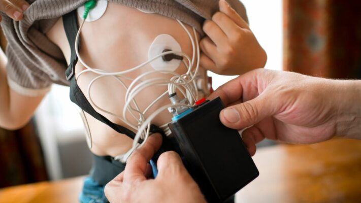
Holter ECG Monitoring in Children: Procedure, Uses and Results
Holter ECG in children is a [...]
Read More
29
Sep
Sep
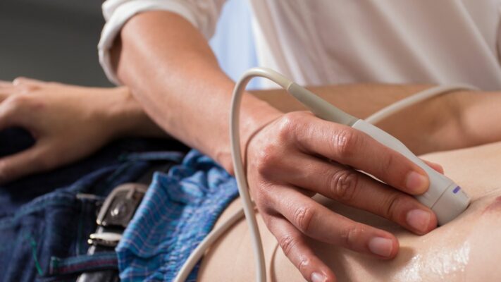
Pediatric Echocardiography (ECHO): Procedure, Uses and Results
Pediatric echocardiography is a painless ultrasound [...]
Read More
29
Sep
Sep
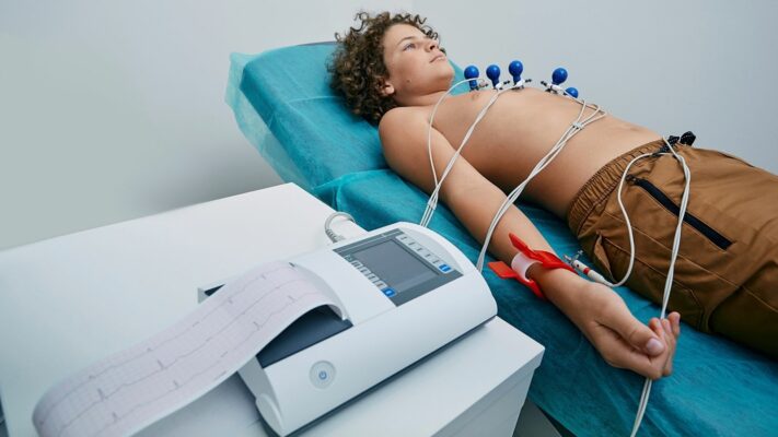
Pediatric ECG (Electrocardiogram): Procedure, Uses and Results
Pediatric ECG (Electrocardiography) is a completely [...]
Read More
29
Sep
Sep

Pulmonary Valve Stenosis: Symptoms, Causes, Diagnosis and Treatment
Pulmonary valve stenosis (pulmonary stenosis) is [...]
Read More
29
Sep
Sep
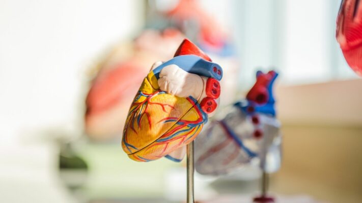
Ventricular Septal Defect (VSD): Symptoms, Causes and Treatment Options
Ventricular septal defect (VSD) is a [...]
Read More
29
Sep
Sep
Atrial Septal Defect (ASD): Symptoms, Causes and Treatment Options
Atrial septal defect (ASD) is a [...]
Read More
29
Sep
Sep

Shortness of Breath in Children: Causes, Symptoms and Treatment Options
Shortness of breath in children is [...]
Read More
10
Aug
Aug