
 Türkçe
 Deutsch
 العربية
 Русский

Inflammation of the pericardium that develops later in children (pericarditis) is the inflammation of the membrane structure surrounding the heart after infection, autoimmune disease, or trauma. This condition presents with symptoms such as chest pain, fever, and fatigue, and requires early diagnosis and appropriate treatment planning.
Symptoms of pericarditis in children are generally seen as sudden-onset chest pain, a stabbing pain that increases with deep breathing, and palpitations. In some patients, fever, cough, and deterioration in general condition may accompany it. Clinical evaluation is supported by auscultation findings, echocardiography, and electrocardiography. In addition, blood tests also guide us in terms of treatment.
Among the causes of pediatric pericarditis, viral infections are in the first place, while bacterial infections, rheumatologic diseases, and inflammation developing after cardiac surgery may also be factors. Correctly determining the cause plays a decisive role in planning the treatment approach.
Treatment of pericarditis in children may include anti-inflammatory medications, antibiotics, or immunosuppressive therapies depending on the underlying cause. In cases where fluid accumulation develops, pericardiocentesis may be required both for diagnosis and for treatment. Regular cardiology follow-up is important in terms of reducing the risk of complications.
| Things You Should Know | Information |
| Definition of the Disease | Inflammation of the pericardium that develops later in children (pericarditis) is characterized by inflammation of the membrane structure surrounding the heart. As a result of inflammation, fluid accumulation may occur in the pericardial space and the heart’s normal filling function may be affected. |
| Function of the Pericardium | The pericardium is the membrane structure that protects the heart from external factors, stabilizes the heart’s position in the chest cavity, and prevents excessive expansion. It also reduces friction during heart movements. |
| Frequency | It is rare in childhood but may occur after infections. It is diagnosed more frequently in school-age children and adolescents. |
| Main Causes | Viral infections are the most common cause. In addition, bacterial infections, tuberculosis, autoimmune diseases, rheumatic diseases, post-cardiac surgery conditions, trauma, and kidney failure may be causes. |
| Risk Factors | A recent upper respiratory tract infection, immune system diseases, chronic kidney disease, history of heart surgery, and systemic infections create risk. |
| Pathophysiology | As a result of inflammation, an increase in fluid occurs between the pericardial layers. Rapid fluid accumulation can prevent the filling of the heart chambers and lead to cardiac tamponade. |
| Symptoms | Chest pain, fever, fatigue, palpitations, shortness of breath, and abdominal pain in young children may be seen. |
| Characteristic of Chest Pain | It is usually sharp and stabbing. It increases with deep breathing, may decrease when leaning forward, and may radiate to the back or shoulder. |
| Physical Examination Findings | A pericardial friction rub may be heard. Tachycardia, low blood pressure, and in advanced cases jugular venous distension may be detected. |
| Cardiac Tamponade | It is the severe impairment of cardiac filling due to increased fluid in the pericardial space. It is characterized by hypotension, tachycardia, and muffled heart sounds and requires URGENT intervention. |
| Diagnostic Methods | Clinical evaluation, electrocardiography (ECG), echocardiography, chest X-ray, and blood tests are used. When necessary, advanced imaging methods may be utilized. |
| ECG Findings | Diffuse ST-segment elevation and PR-segment depression may be seen. In later stages, T-wave changes may occur. |
| Echocardiography | It is the most important imaging method for determining the presence and amount of pericardial fluid. It enables evaluation of cardiac functions. |
| Laboratory Findings | Inflammation markers such as C-reactive protein (CRP) and erythrocyte sedimentation rate (ESR) may be elevated. Tests directed toward infection are performed. |
| Differential Diagnosis | It can be confused with myocarditis, pneumonia, pleuritis, musculoskeletal chest pain, and reflux disease. |
| Treatment Approach | Cause-directed treatment is essential. Mostly nonsteroidal anti-inflammatory drugs and rest are recommended. In bacterial infections, appropriate antibiotic therapy is administered. |
| Advanced Intervention | If cardiac tamponade develops, drainage of fluid with pericardiocentesis may be required. In resistant cases, corticosteroid therapy may be considered. |
| Need for Hospitalization | Hospitalization is required in severe symptoms, presence of large effusion, signs of tamponade, and suspicion of systemic infection. |
| Possible Complications | Cardiac tamponade, recurrent pericarditis, and rarely constrictive pericarditis may develop. |
| Recurrent Pericarditis | It may recur weeks or months after the first attack. It may require long-term follow-up and treatment with different medications. |
| Prognosis | In cases of viral origin, it generally has a good course and completely resolves with appropriate treatment. Prognosis varies depending on the underlying cause. |
| Follow-up | Regular follow-up is performed with clinical findings, inflammation parameters, and echocardiography. |

PROF. DR.
İbrahim Cansaran TANIDIR
Pediatric Cardiology Specialist
İbrahim Cansaran TANIDIR
Pediatric Cardiology Specialist
Prof. Dr. İbrahim Cansaran Tanıdır is an internationally recognized expert with nearly 20 years of clinical, academic, and interventional experience in congenital heart diseases, pediatric interventional cardiology, hybrid procedures, and radiation-reduced / fluoroscopy-free catheterization techniques.
To date, he has performed more than 10,000 pediatric cardiac catheterization procedures both in our country and abroad; and has pioneered advanced interventional and hybrid applications in complex structural heart diseases in our country. His academic productivity represents a strong scientific authority with 110 SCI/SCIE publications, 1,440 citations, and an H-index of 18 (Google Scholar, 2025).
What Causes Pericarditis in Children and Which Factors Play a Role?
When a child who was completely healthy suddenly presents with chest pain and is diagnosed with pericarditis, families are usually surprised. “Why did this happen to us?” is the question we hear most often. In childhood, the biggest culprit of this inflammation is actually viral infections that we know very well. Most of the time, the child has had a mild upper respiratory tract infection, the flu, or a gastrointestinal infection 1–2 weeks before pericarditis develops. The fever has gone down and the child seems to be getting better, but while the immune system is fighting the virus, it may get a little “confused” and mistakenly attack the child’s own pericardium. We call this a “post-viral” reaction, meaning a reaction that develops after a virus. Sometimes the virus itself settles in the pericardium.
Bacterial infections, although much rarer today thanks to the widespread and effective use of antibiotics, are among the most challenging conditions when we encounter them. They usually occur when pneumonia, a severe bone infection, or an infection that has entered the bloodstream spreads to the pericardium. In this case, the fluid that accumulates in the pericardium may not be clear but may become purulent, requiring a much more aggressive treatment.
In addition, non-infectious causes should not be overlooked. Autoimmune diseases, in which the body wages war against its own tissues, can cause pericarditis. Especially in the course of diseases such as Acute Rheumatic Fever, known as joint rheumatism, Juvenile Idiopathic Arthritis, or Lupus (SLE), pericardial involvement may be seen. In children with kidney failure, elevated blood urea levels or similar conditions may also be encountered during the healing process after cardiac surgeries.
The most common viral agents that cause this disease are as follows:
- Coxsackie viruses
- Echoviruses
- Adenoviruses
- Influenza
- Parvovirus B19
- Epstein–Barr virus
Which Symptoms of Pericarditis Should Families Be Alert To?
The most typical and distinctive symptom of pericarditis is chest pain. However, this pain has very different characteristics from any muscle pain or heartburn. School-age children and adolescents who can express themselves usually describe this pain as “like being stabbed” or “sharp.” The most important characteristic of the pain is that its severity changes depending on the child’s posture.
When the child lies on their back or takes a deep breath, the pain can become unbearable. Because in these positions, the inflamed pericardial layers come into greater contact with each other. In contrast, when the child sits leaning forward (which we call the tripod position in medicine), they feel that the pain eases. For this reason, rather than forcing a child with pericarditis to lie down in bed, allowing them to rest in a sitting position provides more comfort. The pain may sometimes not remain only in the middle of the chest; it may radiate to the neck, left shoulder, and the muscles in the upper part of the back. This radiation is due to the nerve relationships of the pericardium.
In infants and young children who cannot yet speak, making a diagnosis depends entirely on the parents’ observation ability and the physician’s suspicion. They cannot say “my chest hurts.” Instead, restlessness, unexplained crying spells, loss of appetite, and moaning may be seen. If your baby has recently had a febrile illness and now calms down when you hold them but starts crying when you lay them on their back in the bed, this can be a very important clue for pericardial inflammation.
When the disease progresses and fluid starts to accumulate between the pericardial layers, shortness of breath, easy fatigue, and sometimes a dry cough may be added to the picture. As the amount of fluid increases, the heart’s ability to fill comfortably is hindered, which can lead to symptoms that mimic signs of heart failure such as abdominal pain or swelling in the legs.
The main symptoms to pay attention to are as follows:
- Sharp chest pain
- Pain that increases when lying on the back
- Pain that decreases when leaning forward
- Pain radiating to the left shoulder
- Fever
- Fatigue
- Shortness of breath
- Sensation of palpitations
- Dry cough
- Loss of appetite
How Is Pericarditis Diagnosed During the Doctor’s Examination?
While examining your child, our most valuable diagnostic tool is our stethoscope. When listening to the heart of a child with pericarditis, we look for a very characteristic sound in addition to normal heart sounds. This sound, which we call a “friction rub” in medicine, occurs when two inflamed and roughened pericardial layers rub against each other with every heartbeat. To describe this sound; we can liken it to the creaking sound when walking on snow or the squeak of a new leather jacket. Hearing this sound almost confirms the diagnosis. However, we may not always be able to hear it. If the amount of fluid accumulated between the layers has increased, the friction sound disappears because the layers move apart. In this case, heart sounds may sound as if they are coming from very far away, “muffled.”
After the physical examination, the first test we use is Electrocardiography, namely the ECG. This simple test, which maps the electrical activity of the heart, shows very typical wave changes in pericarditis. For families who come with concern about a heart attack, showing that these unique changes on the ECG (such as diffuse ST elevations) indicate pericarditis and explaining that this is not a heart attack is reassuring. ECG findings change from day to day depending on the stage of the disease, so during the treatment process we frequently obtain ECGs to monitor recovery.
Blood tests help us understand the extent of inflammation in the body. Infection and inflammation markers such as CRP, sedimentation, and white blood cell values are usually elevated. In addition, the Troponin test is very critical for us. This value, which normally rises in heart muscle damage, may also rise slightly in pericarditis. If troponin is elevated, we understand that the inflammation is not limited to the membrane but has also spread to the muscle layer of the heart (myocardium). This is called “myopericarditis” and requires us to monitor the course a bit more carefully.
To visualize the diagnosis and measure the amount of fluid, the gold standard is Echocardiography (ECHO). With this method, in which we image the heart with ultrasound waves, we can measure thickening of the pericardium and the fluid accumulated between the layers in millimeters. More importantly, we can immediately see whether this fluid is compressing the heart (tamponade effect).
The methods used in the diagnostic process are as follows:
- Physical examination
- Auscultation with a stethoscope
- Electrocardiography
- Echocardiography
- Chest X-ray
- Complete blood count
- CRP test
- Sedimentation test
- Troponin test
- Computed tomography
- Magnetic resonance imaging
Contact us now to get detailed information about treatments and to create an appointment!
Which Medications and Methods Are Used in the Treatment of Pericarditis?
When we diagnose pericarditis, the family’s first question is, “Will you prescribe antibiotics?” However, as mentioned above, the most common cause of pericarditis in children is viruses, and antibiotics do not work against viruses. Therefore, if we are not sure of a bacterial infection, antibiotics have no place in treatment. The main goals of our treatment are to relieve pain, resolve inflammation, and most importantly prevent recurrence (relapse) of the disease.
The backbone of treatment is “Nonsteroidal Anti-Inflammatory Drugs” (NSAIDs). Ibuprofen is the medication we most frequently prefer in this group, with proven safety in children. However, there is a very important nuance here: We use this medication not only at analgesic or antipyretic doses, but at high “anti-inflammatory” doses that suppress inflammation. The most common warning we give families is not to fall into the mistake of saying, “The child’s pain is gone, let’s stop the medication.” Even if the pain resolves, microscopic inflammation may still persist. Therefore, the medications should be used regularly on schedule for the duration determined by the physician (usually a few weeks) and should be tapered and discontinued slowly.
Another important point to consider here is whether it is accompanied by myocarditis. Because the medications to be given differ between these two disease groups.
The second hero of treatment is “Colchicine.” This old medication, known for Familial Mediterranean Fever or gout, has changed the rules of the game in pericarditis treatment. Scientific studies have shown that when low-dose colchicine is added to NSAID therapy, the rate of recovery increases and the risk of recurrence decreases by half. For this reason, we definitely add it to our treatment protocol especially in the first attack and in recurrent cases. It is generally used for a long period such as 3 months.
The use of cortisone (steroids) is the most debated issue in pediatric pericarditis. Cortisone is a very strong anti-inflammatory and corrects symptoms rapidly, sometimes within a day. This can be tempting for families and the physician. However, the other side of the coin is different. Using cortisone in viral pericarditis may delay clearance of the virus from the body and increase the likelihood that the disease will return “like a boomerang” when the medication is stopped. Therefore, unless there is an autoimmune disease or a special condition, we avoid cortisone as much as possible in first-line treatment. If we have to use it, we start at very low doses and taper it very slowly over months.
The main medication groups used in treatment are as follows:
- Ibuprofen
- Naproxen
- Indomethacin
- Aspirin
- Colchicine
- Prednisolone
- Anakinra
- Gastric protectants
What Is Done If Pericardial Fluid Increases and What Is Tamponade?
In most pericarditis cases, a small amount of fluid accumulates between the layers, and this fluid is gradually absorbed by the body along with medication treatment. However, sometimes things may not go as we want. The fluid accumulates very rapidly or the amount increases so much that the heart cannot withstand this external water pressure. The heart is a pump that needs to fill with blood every time it relaxes. If it is surrounded by pressurized fluid, it cannot expand sufficiently and cannot take in enough blood. A heart that cannot take in blood cannot pump blood to the body. Blood pressure drops, the pulse speeds up but becomes weak, and the child deteriorates. We call this condition “Cardiac Tamponade,” and it is a life-threatening situation requiring urgent intervention.
When tamponade develops, we do not have time to wait for medications to take effect. We must mechanically remove this pressure on the heart. This procedure is called “pericardiocentesis.” Usually under ultrasound (ECHO) guidance, a special needle is inserted through the chest wall (generally just below the sternum) and the excess fluid between the pericardial layers is drained. The moment the needle enters and the fluid begins to drain, the child’s blood pressure improves, their color returns, and they feel relief. This procedure is both life-saving and diagnostic. Because we can send the fluid we obtain to the laboratory to examine whether it contains bacteria, tuberculosis germs, or tumor cells.
If the fluid is very thick, purulent (purulent pericarditis), or recurs continuously, drainage with a needle alone may not be sufficient. In this case, it may be necessary to surgically place a tube through a small incision to allow drainage of the fluid, or to create a “window” in the pericardium so that the fluid can drain into the abdominal cavity or the chest cavity.
Situations requiring urgent intervention are as follows:
- Low blood pressure
- Shock picture
- Cardiac tamponade
- Presence of purulent fluid
- Lack of response to treatment
- Diagnostic uncertainty
- Suspicion of tumor
Contact us now to get detailed information about treatments and to create an appointment!
Can Pericarditis Become Chronic and Leave Permanent Damage?
The biggest fear of families is whether this disease will leave permanent heart damage. The good news is that the vast majority of children who have acute viral pericarditis recover completely and continue their lives without any restrictions. The pericardium is a structure that can repair itself. However, there are exceptions to every rule. Especially chronic infections such as tuberculosis, untreated bacterial pericarditis, or continuously recurring attacks may lead to permanent changes in the pericardium.
If the inflammatory process lasts for a very long time, the pericardium may lose its elasticity and become thickened, hardened, and even calcified. In medicine, we call this “Constrictive Pericarditis.” The membrane surrounding the heart is no longer like an elastic balloon but like a hard armor or eggshell. Even if the heart muscle is intact, it cannot expand within this armor. This condition can cause fluid accumulation in the child’s abdomen, edema in the legs, and a significant decrease in exercise capacity.
When constrictive pericarditis develops, unfortunately, medication treatments cannot restore this hardened tissue to its former state. The only solution is surgery. With an operation called “Pericardiectomy,” this thickened membrane that constricts the heart is peeled off and removed. After surgery, the heart regains its freedom and the child’s complaints improve dramatically. Fortunately, we see this condition quite rarely in children, but the main reason not to neglect follow-up is to detect this risk early.
Symptoms of constrictive pericarditis are as follows:
- Abdominal swelling
- Edema in the legs
- Fullness in the neck veins
- Easy fatigue
- Enlargement of the liver
- Loss of appetite
- Weight loss
What Should Families Pay Attention To During the Recovery Process and How Long Should Sports Restrictions Last?
Being discharged from the hospital or starting medication treatment does not mean the process is over. In fact, the home care process is at least as critical as medications for the success of the treatment. For a child who has had pericarditis, “rest” is not just a recommendation but a part of the prescription. In the acute period, that is, while the child has pain and fever, absolute bed rest is essential. Because as the heart rate increases, the number of friction events increases and the healing tissue becomes irritated again.
The issue that challenges families the most is keeping a child who feels well inactive. The answer to the question “I feel fine, can I go out and play ball?” must unfortunately be “No” for a while. Our general rule is this: If there is only pericarditis (the heart muscle is not affected), even after blood values (CRP) and ECG have completely returned to normal, competitive sports should be avoided for at least 3 months. If there is myopericarditis (heart muscle involvement), this period extends to at least 6 months. During this period, the child can go to school and perform daily activities, but should not participate in physical education classes, should not carry a heavy backpack, and should avoid training that would strain the heart.
The decision to return to sports should not be made by the family but 반드시 by a pediatric cardiologist. After ECG, ECHO, and if necessary an exercise test or Holter are performed at the follow-up examination, the “green light” is given.
The follow-up process is also long-term. Even after treatment ends and medications are discontinued, the risk of recurrence in the first year is between 15–30%. For this reason, it is necessary to go for check-ups at certain intervals even if there are no complaints. Families should consult a doctor without waiting for the appointment date if chest pain recurs, sudden fever rises, or the child cannot exert as much effort as before. Pericarditis is a process that requires patience; however, with the right treatment, good rest, and regular follow-up, children can recover without leaving a trace on their heart.
Things to pay attention to during the recovery period are as follows:
- Bed rest
- Avoiding strenuous exercise
- Not carrying a school bag
- Regular medication use
- Plenty of fluid intake
- Balanced nutrition
- Regular doctor follow-up
- Fever monitoring
- Avoiding stress
Frequently Asked Questions
After which infections does inflammation of the pericardium that develops later in children (pericarditis) most commonly occur?
In children, pericarditis most commonly develops after viral upper respiratory tract infections. Rarely, bacterial infections, rheumatic diseases, or immune system problems may also trigger inflammation in the pericardium.
Is this condition more severe in children with immune system diseases?
In children with autoimmune disease, the immune system may mount an excessive response against the pericardium. This can cause the inflammation to be more severe and recurrent, therefore close follow-up and regular monitoring are important.
What risks does fluid accumulation in the heart pose in children who have had pericarditis?
If fluid accumulation in the pericardium increases, it can prevent the heart from filling comfortably. This condition is known as cardiac tamponade and requires urgent intervention. With early diagnosis, serious complications can largely be prevented.
How is pericarditis pain distinguished in children experiencing chest pain?
Pericarditis pain is usually a stabbing chest pain that decreases when leaning forward and increases when lying on the back. It may worsen with deep breathing. These features help differentiate it from muscle pain or simple infections. However, definitive differentiation can be made with ECG and echocardiography.
Does pericarditis that develops later in children recur?
In some children, especially viral- or autoimmune-related pericarditis may recur. Appropriate medication treatment and regular follow-up reduce the risk of recurrence. In recurrent cases, longer-term treatment can be planned.
How soon can children diagnosed with pericarditis return to normal activities?
In mild cases, response to treatment is usually seen within a few weeks. However, for sports and intense physical activities, doctor approval should be awaited. Early exertion can prolong the recovery process.
In which situations is surgical intervention necessary?
If increasing fluid accumulation or cardiac compression develops despite medication treatment, the fluid can be drained with a procedure called pericardiocentesis. Very rarely, surgical removal of the thickened membrane may be required.
Does pericarditis cause permanent heart damage in children?
With early diagnosis and appropriate treatment, most children recover completely. However, in untreated or severe cases, thickening of the pericardium may develop and this may affect heart functions.
How can this disease affect children’s psychology?
Chest pain and the hospital process can create anxiety in children. Especially in recurrent attacks, sports restrictions can create social effects. Family support and proper information have a positive effect on the process.
Why is long-term follow-up important after pericarditis?
Even if recovery is achieved, regular cardiology follow-ups are recommended against the risk of recurrence or late complications. A safe recovery process is ensured by monitoring cardiac functions with ECG and echocardiography.
Blog Posts

Exercise Stress Test in Children: Purpose, Procedure and Results
Exercise stress testing in children is [...]
Read More
29
Sep
Sep
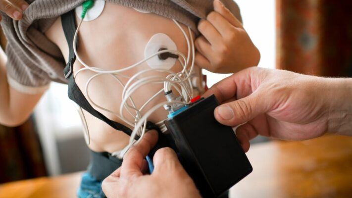
Holter ECG Monitoring in Children: Procedure, Uses and Results
Holter ECG in children is a [...]
Read More
29
Sep
Sep
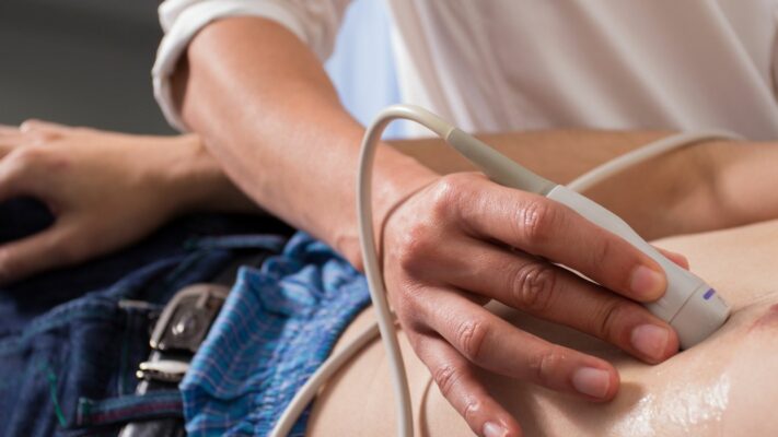
Pediatric Echocardiography (ECHO): Procedure, Uses and Results
Pediatric echocardiography is a painless ultrasound [...]
Read More
29
Sep
Sep
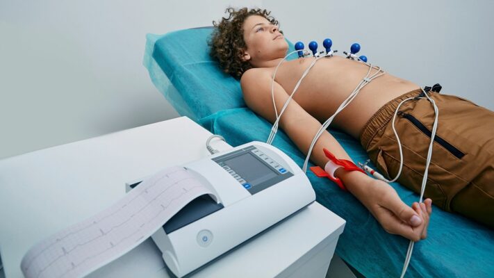
Pediatric ECG (Electrocardiogram): Procedure, Uses and Results
Pediatric ECG (Electrocardiography) is a completely [...]
Read More
29
Sep
Sep

Pulmonary Valve Stenosis: Symptoms, Causes, Diagnosis and Treatment
Pulmonary valve stenosis (pulmonary stenosis) is [...]
Read More
29
Sep
Sep
Ventricular Septal Defect (VSD): Symptoms, Causes and Treatment Options
Ventricular septal defect (VSD) is a [...]
Read More
29
Sep
Sep
Atrial Septal Defect (ASD): Symptoms, Causes and Treatment Options
Atrial septal defect (ASD) is a [...]
Read More
29
Sep
Sep

Shortness of Breath in Children: Causes, Symptoms and Treatment Options
Shortness of breath in children is [...]
Read More
10
Aug
Aug